Diffusion in Agar Cubes

Hypothesis

  • Smaller the cube is, faster it is diffuse as it has smaller surface area.

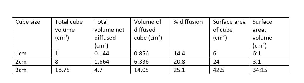
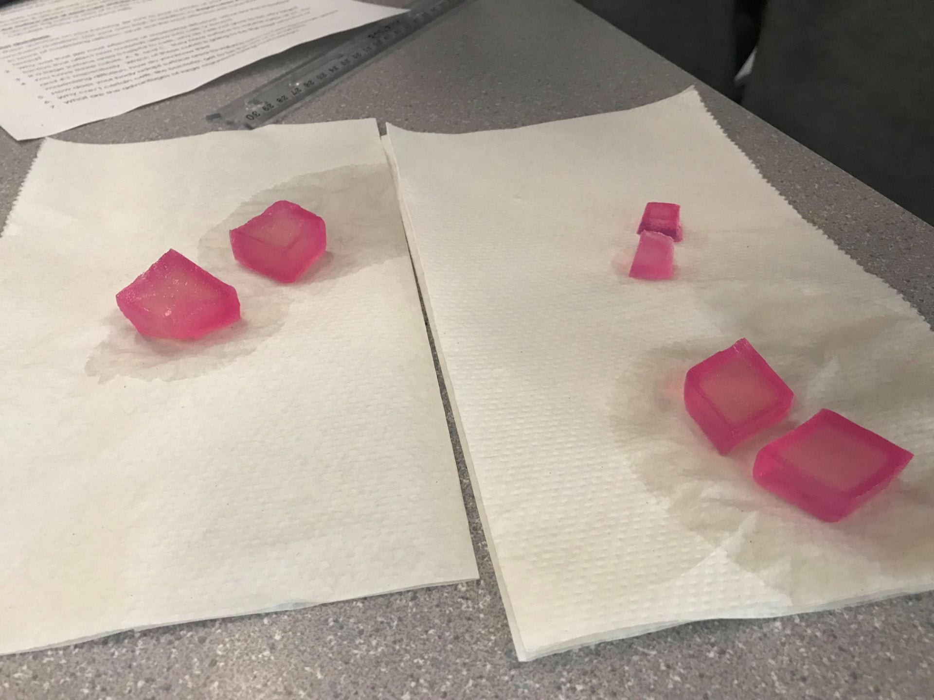
In terms of maximizing diffusion, what was the most effective size cube that you tested?

  • The 1cm cube was the most effective one.

Why was that size most effective at maximizing diffusion? What are the important factors that affect how materials diffuse into cells or tissues?

  • The smaller the size, higher the SA: V ratio. Due to small surface area, it has less surface to cover. Therefore, could have many materials pass through in and out of the “cell” by diffusion.
  • Some important factors might be time, size, and possibly temperature.

If a large surface area is helpful to cells, why do cells not grow to be very large?

  • Cells would not grow bigger as it would be difficult to transport nutrients to the center and would take longer to regenerate. However, smaller the size, the faster it would be to travel and take lesser time overall.

You have three cubes A, B, and C. They have surface to volume ratios of 3:1, 5:2, and 4:1 respectively. Which of these cubes is going to be the most effective at maximizing diffusion, how do you know this?

  • C has the highest surface area to volume ratio. This means that for every cubic unit of cytoplasm, there are more cell membrane than in cubes A and B. This allows for more materials to be able to enter the cytoplasm through diffusion. Therefore, cube C will be most effective at maximizing diffusion.

How does your body adapt surface area to volume ratios to help exchange gases

  • Our body adapts the ratio for Alveoli, an air-filled sac inside our lungs. They need a large surface area to volume ratio to allow gas exchange to occur more rapidly in our body.

Why can’t certain cells, like bacteria, get to be the size of a small fish?

  • Bacteria are single-celled organisms. As seen in the lab, cells prefer to have a lower volume, so being the size of a fish would not help the functions of the cell. A cell needs to stay small for diffusion of materials.

What are the advantage of large organisms being multi-cellular?

  • The advantages are that protein can enter into cells directly, rather then needing the assistance of materials to diffuse into the center of a bigger cell.

DNA and Protein Synthesis – Transcription and Translation

Transcription

How is mRNA different than DNA?

The function of mRNA is slightly different than DNA – mRNA copies the information needed to build a protein, which is located on one gene of DNA. mRNA then transfers to the cytoplasm where the protein is built. mRNA is also shorter than DNA and mRNA has one strand while DNA has two

Describe the process of transcription.

Transcription is the process of bringing instructions to build a protein from DNA in the nucleus to ribosomes in the cytoplasm. This is done by messenger RNA (mRNA). In the nucleus, the DNA unzips itself at the location of the gene that needs to be read. This is called the sense strand of the gene and is shown by the blue pipecleaner strand. The mRNA then builds a copy using complimentary base pairs of the strand, which is the red pipecleaner strand in the photo. Blue Thymine beads also change to brown Uracil beads on the mRNA strand. This is all facilitated by the RNA polymerase, or the fuzzy peach in the photo. It separates itself from the DNA when the gene copy is made and the RNA transfers out of the nucleus and into the cytoplasm. The mRNA is then read by ribosomes in the cytoplasm to build the protein in the translation step.

How did today’s activity do a good job of modelling the process of RNA transcription? In what ways was our model inaccurate?

Today’s activity did a good job modelling RNA transcription because we had a different color bead to represent Uracil from Thymine, a different colored pipecleaner for the RNA strand, and the pipecleaners make it easy to separate the nitrogen bases bonds. One way our model was inaccurate was that since our DNA strand was very short, the mRNA made a copy of the whole DNA, calling the whole strand a “gene”. This is possible, but a gene is usually just a section of DNA. In the body, the DNA only unwinds in that specific area, the mRNA makes a copy of the gene, and then the DNA reforms whole again. As well, RNA strands are usually shorter than DNA strands, but in this activity, they were the same size.

Translations Describe the process of translation: initiation, elongation, termination.

The process of translation is in three steps. The first is initiation. In this step, the ribosome looks for the start codon “AUG” on the mRNA. When it finds this codon, the two ribosome subunits (red paper shape in photo) bind together and start reading the mRNA with AUG in the P site. The second step is elongation. The ribosome brings in tRNA (green paper in photos) with the anticodon that matches codons on the mRNA. The tRNA is also holding the matching amino acid to the codon. The amino acids are the blue papers in the photo. When tRNA binds to the “P” site, another tRNA binds to the codon in the “A” site. When both spots are full, the amino acid detaches from the tRNA on the “P” site and attaches itself to the amino acid on the “A” site. The tRNA in the P site then detaches itself from the ribosome, and the ribosome then moves so that the tRNA in the A site is now in the P site. A new tRNA attaches itself to the new exposed codon in the A site, and the process continues. The last step is termination. The elongation cycle ends when the ribosome reads a “STOP” codon. This is a codon that has no matching tRNA amino acid. No amino acid is added to the chain, so the polypeptide is released, and the ribosome detaches from the mRNA.

How did today’s activity do a good job of modelling the process of translation? In what ways was our model inaccurate?

Today’s activity did a good job modelling the process of translation because the paper made it easy to visualize the process. Because the paper is easy to move, you can model the elongation steps in a very similar way to how it’s done in the body. The matching up codons to anticodons and amino acids was also very easy with these models. One way that the model was inaccurate was that the RNA was missing the phosphate – sugar strand. It only showed the bases. As well, the ribosome was one piece when we started, but the ribosome should be in two subunits and then join during initiation. The shapes of the amino acids on the paper were also not accurate to how they look. They are much more randomly shaped than the paper shows.


DNA (Deoxyribonucleic acid)

Structure of a DNA (Deoxyribonucleic acid)

The structure of DNA is made up of 3 main components. Sugar and phosphate groups making up the 2 “backbones” and nitrogenous bases. The sugar-phosphate on each “backbone” are antiparallel structured, meaning that the phosphates on one “backbone’ reads in one direction and are above the sugar group, while the other “backbone” reads in the opposite direction and the phosphate is below the sugar group. The nitrogenous bases are attached to the sugar group and another nitrogenous base through H-bonding. Each nitrogenous base has a complimentary base pairing. If Guanine (G) is present on one “backbone”, then Cytosine (C) must be connected on the other “backbone”. Same thing happens with Thymine (T) and Adenine (A).

(Section of the nucleotide H-Bonded with their complimentary base)

(Pink  = Phosphate, Yellow = Adenine (A), Blue = Thymine (T), Green = Cytosine (C), Purple = Guanine (G), Blue pipecleaner = “backbones”, White pipecleaner = hydrogen bonds)

How does this activity help model the structure of DNA? What changes could we make to improve the accuracy of the model?

This activity helps understand the basics of modeling a DNA molecule down to larger pieces, which means that showing all bonds within the molecule aren’t necessary. That makes the activity less tedious and time consuming. A way to improve this activity would be to expand the size of the DNA molecule in order to show how the sugar bases, nitrogenous bases and phosphate groups are bonded together. This can be shown by creating each section of a nucleotide separately, then combining them with a couple other nucleotide together to make a “zoomed in” model of a DNA molecule.

DNA replication

When does DNA replication occur?

DNA replication happens when cell occurs. It will allow all new cells to have the same DNA across all the new and old cells.

Name and describe 3 steps involved DNA replication. Why does the process occur differently on the “leading” and “lagging” strands?

  • Unwinding and Unzipping: The double helix DNA molecule is unwound into a anti-parallel strands, then unzipped by the enzyme “DNA helicase” from the bottom of the molecule.

  • Complementary Base Pairing: Next, the enzyme DNA polymerase attaches to a leading strand and a lagging strand, one for each. The leading strand is read from “bottom to top”, 3′ to 5′, meaning that the polymerase would pair the complementary bases to one another constantly until the entire molecule is paired. The lagging strand is read from “top to bottom”, 5′ to 3′, meaning the polymerase would pair complementary bases in sections in order to pair up all nucleotide in the molecule, since the helicase is constantly unzipping more nucleotide behind the lagging polymerase.

  • Joining: The nucleotide on the new strands covalently bond to it’s new partner via DNA ligase. After all nucleotide on both the leading and lagging strands are bonded, the 2 new daughter molecules are successfully duplicated and are wound back up into the double helix shape.

Since each “backbone” can only be read in one direction, the DNA polymerase can only read that strand in the direction it’s given. One DNA polymerase would read towards the helicase where the DNA is being unzipped, while the other DNA polymerase would read away from it, meaning after a certain point, it would have to travel backwards to facilitate the complementary base pairings to the newly unzipped nucleotide.

What did you do to model the complimentary base pairing and joining of adjacent nucleotide steps of DNA replication. In what ways was this activity well suited to showing this process? In what ways was it inaccurate?

We had the blue bigfoot candies represent a DNA polymerase, red bigfoot represent DNA ligase, and a watermelon slice represent the helicase. The blue bigfoot sat on top of the DNA molecule to show that it undergoes pairing. While the red bigfoot sat on top of the DNA molecule as well showing the joining of the complementary base pairs.

(Original DNA with replicated DNA)

This model helps accurately show the basics of what happens to DNA as is undergoes replication. The candies help show an introduction to where the specific enzymes sit as they do their jobs.

The model doesn’t show how the enzymes do their jobs when DNA replication occurs.


Desmos Art Functions Card 2018

https://www.desmos.com/calculator/apfoy7m8oj

It was challenging at first where to start but first thing’s first I set up a foundation anywhere where it’ll be perfect. Second, I think of equations to use to make this face. I did not want to make it tedious but simple and AHA! Quadratic and Linear equations would be a simple one! So, I used them and got a face that I was thinking of and used the same method to cite my name on the grid. At last, went to google image to search for the perfect image where it could be the perfect background to display the work I have created.

Thank you 🙂