How is mRNA different than DNA?
mRNA and DNA have many differences. Firstly, the bases used in DNA and mRNA are different. DNA uses cytosine, thymine, adenine, and guanine, whereas mRNA uses uracil instead of thymine. Another one of the structural differences between mRNA and DNA is that DNA has two backbones and mRNA has one backbone. Since RNA has one backbone it has a different shape than DNA, DNA has a double helix and RNA is untwisted. The backbone of mRNA is composed of ribose and phosphate, and DNA’s backbones are made of deoxyribose and phosphate. mRNA is also much shorter than DNA, it is roughly 1000 nucleotides in length and DNA is around 85,000,000 nucleotides in length.
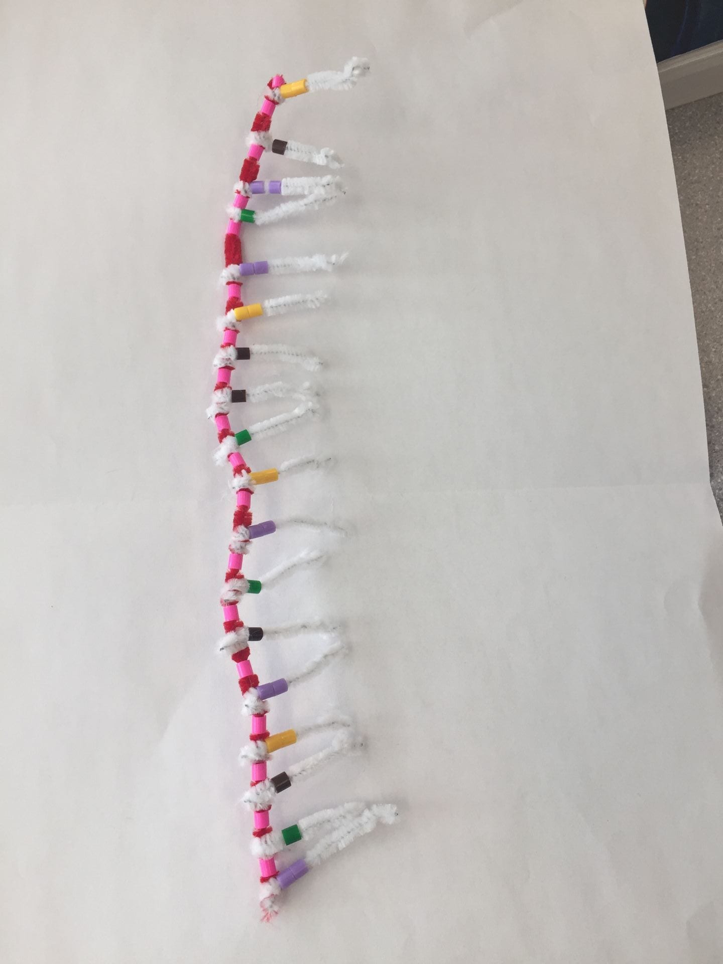
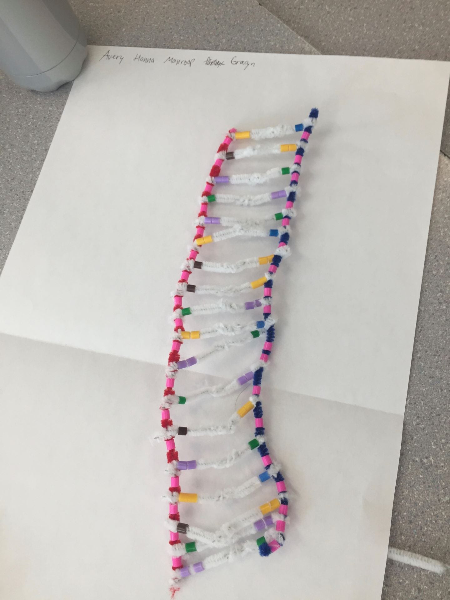
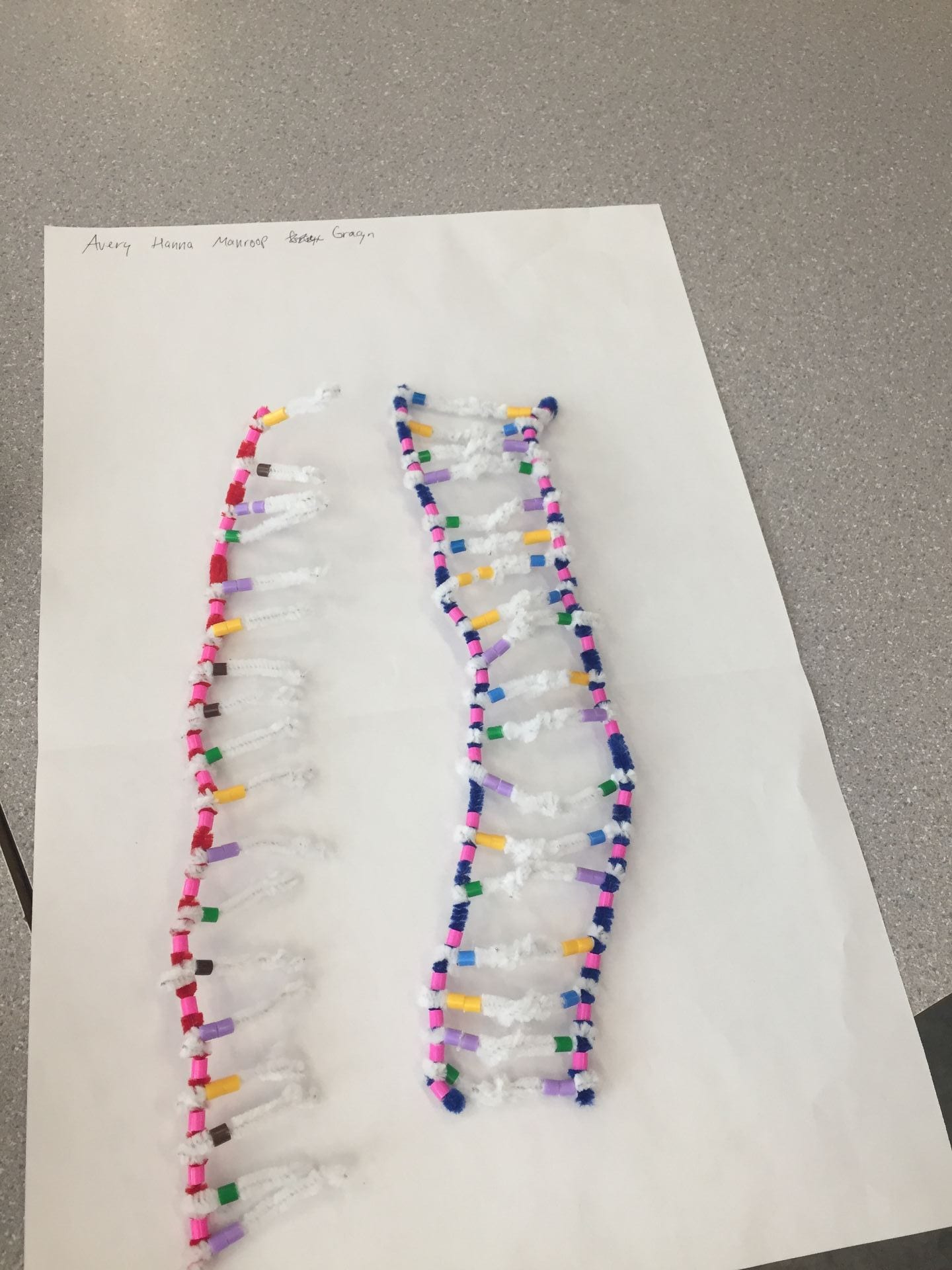
In the pictures below, you can see a side by side comparison of DNA and RNA. DNA is twisted into a double helix, has two blue backbones (deoxyribose), and has blue beads (to represent thymine). RNA is in a straight line, has one red backbone (ribose), and has brown beads (uracil base).

Describe the process of transcription:
The process of transcription happens when mRNA copies the information carried by a gene that is on DNA. Transcription starts in the nucleus and produces mRNA. The process of transcription contains three different steps: unwinding and unzipping DNA, complementary base pairing with DNA, and the separation from DNA. These three steps all happen with the assistance of the enzyme RNA polymerase.
Unwinding:
The first step of transcription is the unwinding of the DNA backbones. The DNA backbones unwind in order for mRNA to copy the information on the DNA strand. The enzyme RNA polymerase unzips DNA at the location of a gene.
Complementary Base Pairing:
In the complementary base pairing step, one strand of DNA is used as a template to build the mRNA. This strand is referred to as the sense strand because it has the instructions for building a protein, whereas the other strand does not have the information needed to build a protein and is not needed in the process of making mRNA, this strand is referred to as the nonsense strand. RNA polymerase places the correct bases on the mRNA strand. Since we are dealing with mRNA, instead of using the base thymine we use the base uracil. This means that adenine pairs with uracil and guanine still pairs with cytosine.
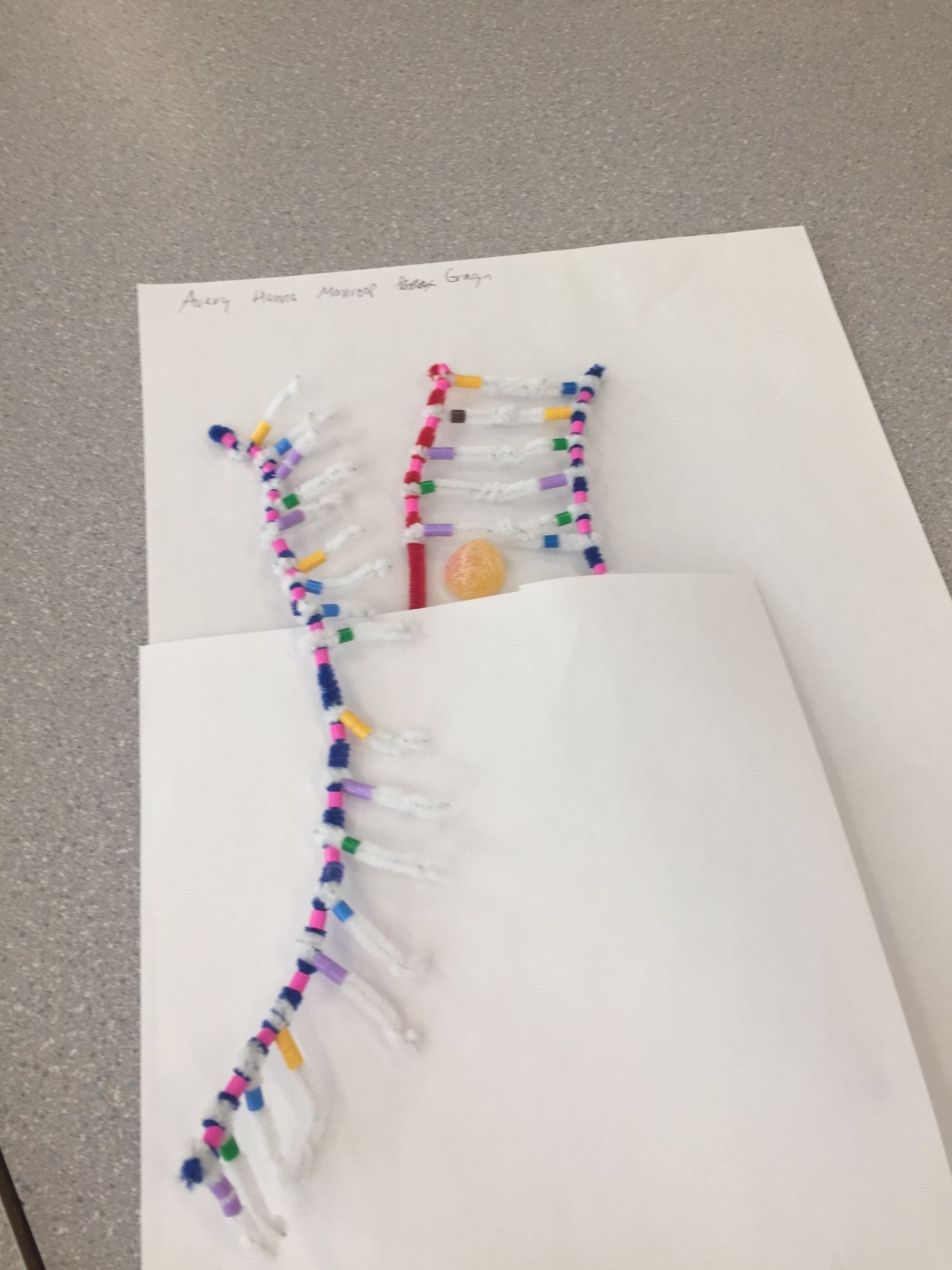
The picture below shows the complementary base pairing step in transcription. The fuzzy peach represents the enzyme RNA polymerase. The mRNA strand is being built using the DNA strand on the right, the sense strand, as a template. The strand on the left is the nonsense strand which is why the mRNA strand is not facing it. We tried to show that RNA polymerase is putting the complementary bases on the mRNA strand by placing the fuzzy peach in the middle of the sense strand and the mRNA strand.
Separation:
Once all a genes information has been copied, the mRNA then separates from the DNA sense strand. The DNA backbones then join together again and twist back into a double helix shape. Although the mRNA has been separated from the DNA strand, it is still modified. After the separation from DNA, the mRNA strand removes sections that do not contain the proper instructions to build a protein. After this modification occurs, the mRNA molecule leaves the nucleus.
How did today’s activity do a good job of modelling the process of RNA transcription? In what ways was our model inaccurate?
I think that today’s activity did a good job representing the differences of RNA and DNA. We were clearly able to see how DNA and RNA differ, like their backbones and the bases. We used blue pipecleaners to represent the deoxyribose backbone along with pink beads to represent the phosphate units on the DNA backbone, whereas for RNA we used red pipecleaners to represent the ribose sugar backbone but still used the pink beads to indicate the phosphate units on the RNA backbone because DNA and RNA have the same type of phosphate units. We also used a different coloured bead on our RNA model to indicate it contained Uracil instead of Thymine. In this activity we continued to use two beads when indicating guanine and adenine, to show that they are double-ringed bases. One of the things that we could have improved was the size of the RNA model. The RNA model was around the same size as the DNA molecule which is inaccurate since DNA is much longer than RNA. We also didn’t go over the process after separation like the modification of mRNA before it leaves the nucleus.
Part II:
Describe the process of translation: initiation, elongation, and termination.
Translation is the process that happens after transcription. In translation, the code carried by mRNA turns into a polypeptide by the help of ribosomes. A ribosome reads the mRNA message and puts amino acids in order and make the protein. The three steps of translation are initiation, elongation, and termination.
Initiation:
The first step of translation is initiation. In this step mRNA is held by a ribosome, which then binds to another ribosome subunit. The ribosome finds the codonAUG in the P-Site on the mRNA strand because this codon initiates the rest of the order of amino acids. The AUG codon pairs with the anticodon UAC. The anticodons can be found by using the complementary base pairs, AUG pairs with UAC because Adenine pairs with Uracil and Guanine pairs with Cytosine.
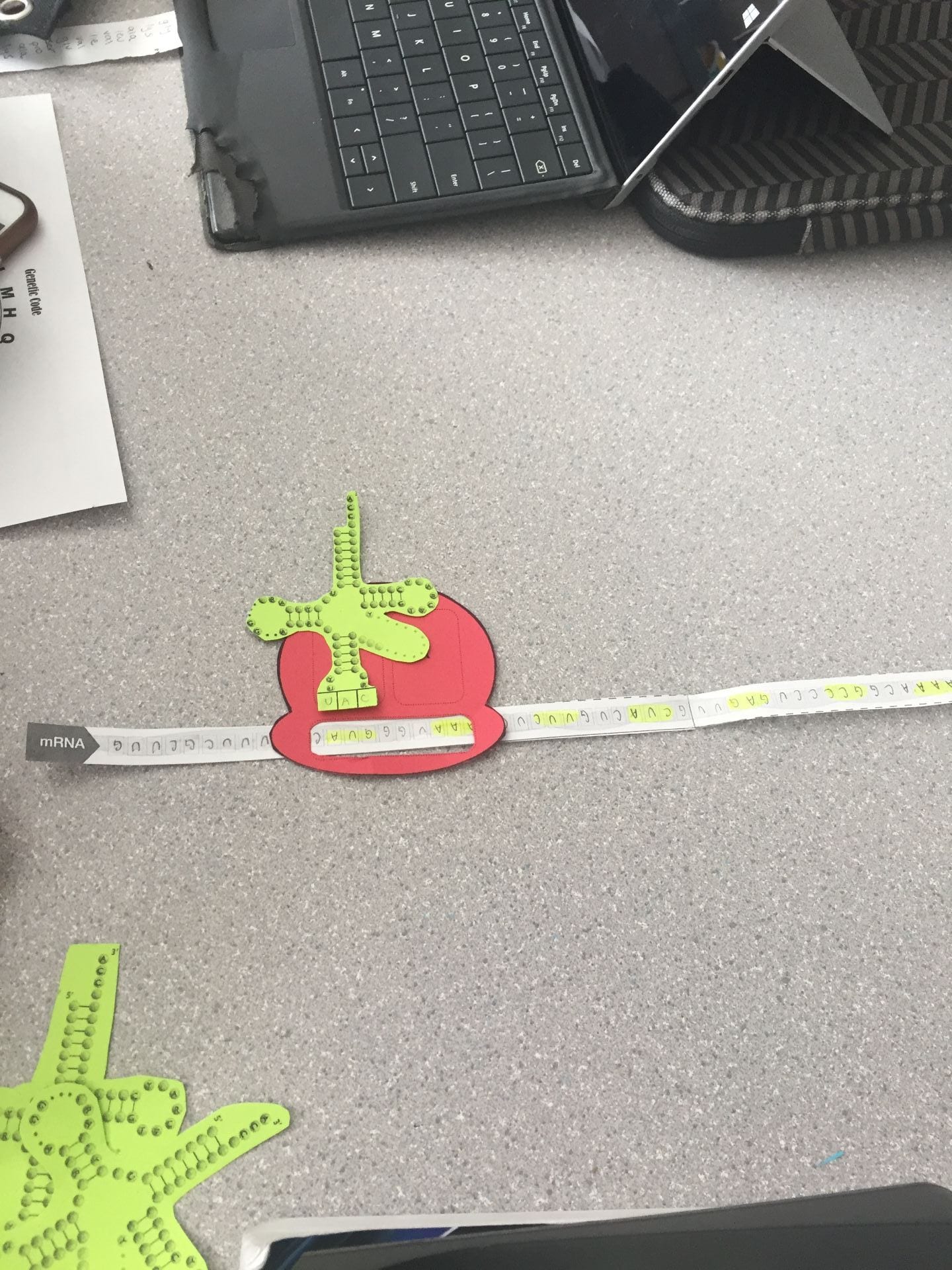
In the picture below, you can see that the ribosome (red piece of paper) goes down the mRNA until it finds the codon AUG. After it finds the codon AUG the anticodon UAC pairs with it which is written on the green piece of paper (the tRNA). Since each codon represents an amino acid, the codon AUG corresponds to the amino acid Methionine which is shown by the blue piece of paper which is also attached to the tRNA.

Elongation:
After the codon AUG is found, the ribosome continues to go down the mRNA strand and reads the codons on it. Codons are three lettered words that indicate specific amino acids and adds another tRNA molecule. Since the first codon AUG is in the P-site, the second codon has to be carried to the A-site. When both the A-site and the P-site are filled, the amino acid at the P-site transfers to the amino acid on the A-site. The tRNA at the P-site floats away once the codon is transferred. The ribosome then moves down the mRNA strand to the next codon which moves to the A-site as the second codon moves to the P-site. The chain of amino acids grows depending on the number of codons using the same process. The corresponding anticodons will also continue to be added to the chain as well. These amino acids keep on binding together which creates a polypeptide chain, the beginning of a protein. This continues on occurring until the tRNA reaches the STOP codon in the A-site.
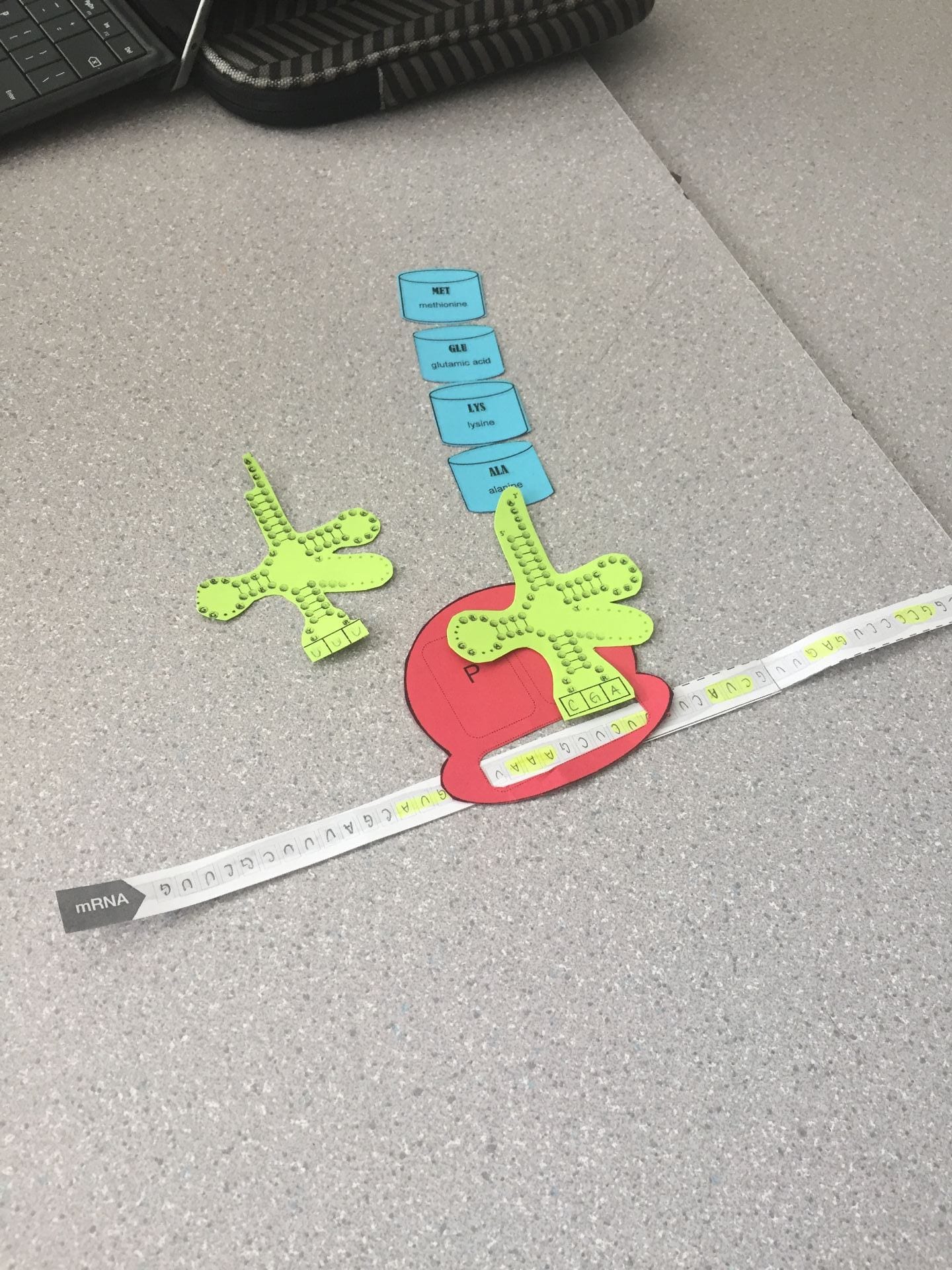
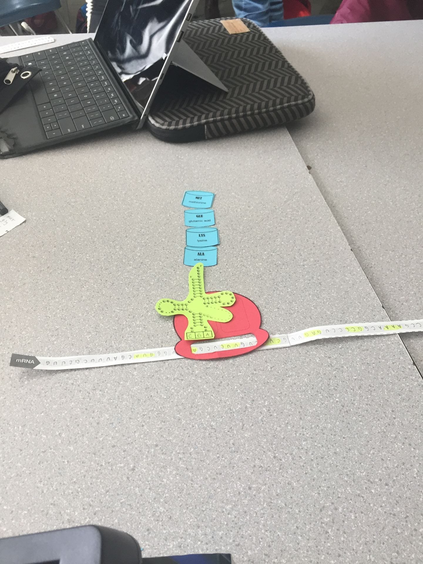
In the picture below, the next codon is read after AUG. The new codon is in the A-site:

The tRNA molecule floats away as the amino acid moves to the A-site:

The ribosome moves down the mRNA strand and the tRNA move back to the P-site:

The process continues to occur:

Termination:
The last step of translation is termination. This is when a stop codon is found, a codon that does not have a matching tRNA. This means that no new amino acid is added to the chain which causes the polypeptide to be released with the use of hydrolysis. The ribosome are released and also split back to their subunits.
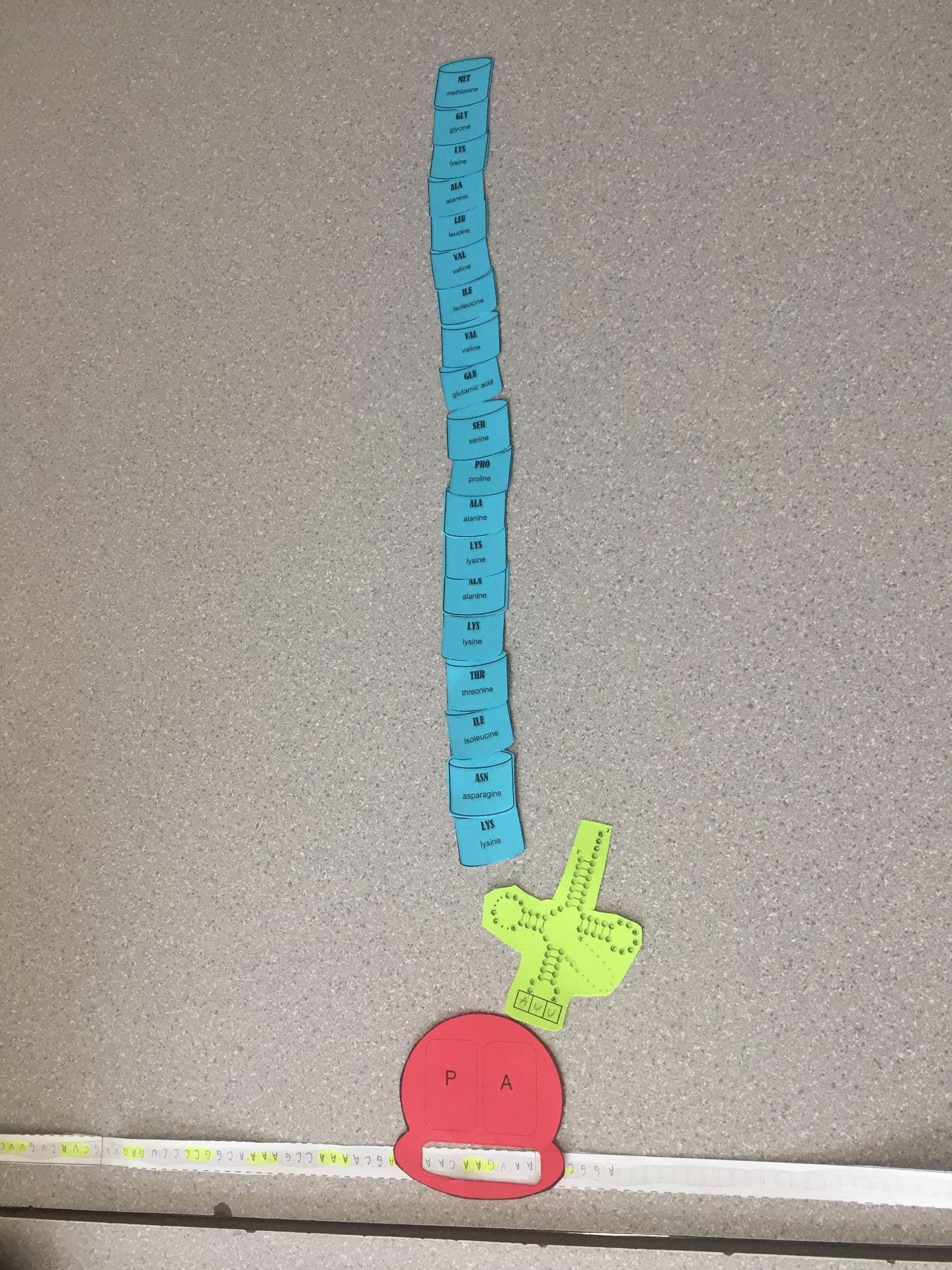
This picture shows our final amino acid sequence. Our amino acid sequence matched to the Topoisomerase protein:

Then the polypeptide amino acid chain is released from the tRNA and ribosome:
How did today’s activity do a good job of modelling the process of translation? In what ways was our model inaccurate?
This activity really helped me understand the process of translation because at we were able to visualize it in an organized way. I liked how the different components involved in the process were separate colours, like the ribosome was red and the tRNA was green. One thing that was inaccurate was that there weren’t the subunits needed for the ribosome, we just had one large ribosome, this also effected the termination step because we weren’t able to show the ribosome breaking down. Another inaccurate factor was the shapes of the amino acids, on the protein sheet it shows that each amino acid has a unique shape but for this activity all of them were the same shape.