Anatomy & Physiology – DNA Replication & Protein Synthesis
Replication:
1. Explain the structure of DNA – use the terms nucleotides, antiparallel strands, and complementary base pairing.
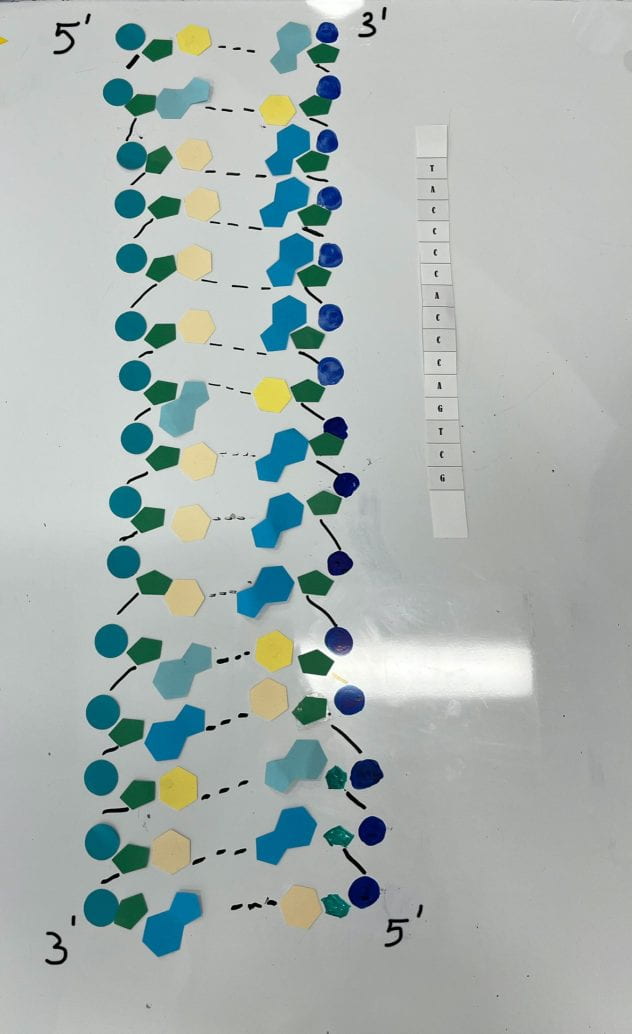
DNA is a polymer composed of nucleotides. The structure of DNA is a double helix formed between antiparallel strands, which means that one strand runs from the 5’ to 3’ end and the opposing strand goes from the 3’ to 5’ end. The two strands are held together by hydrogen bonds that form between complementary bases. (A-T, G-C) The backbone is composed of a 5-carbon deoxyribose sugar covalently bonded with the phosphate group.
2. When does DNA replication occur?
DNA undergoes replication before the cell divides. This ensures that the daughter cells have an identical copy of the DNA in the nucleus. Replication also results in two identical DNA molecules that have been semi-conserved.
3. Name and describe the 3 steps involved in DNA replication. Why does the process occur differently on the “leading” and “lagging” strands?
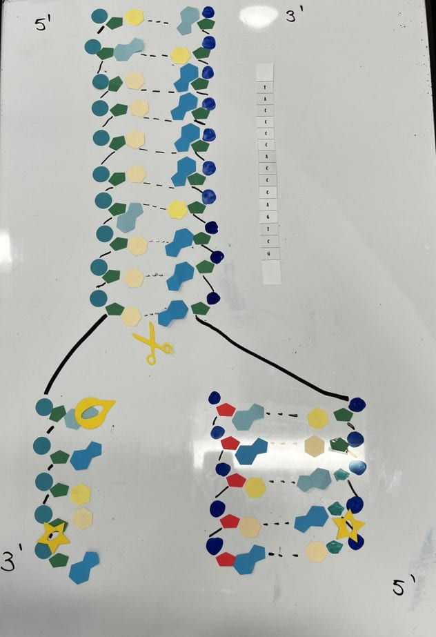
The first step in replication is the unzipping of the DNA. The DNA helicase breaks the hydrogen bonds between the nitrogenous bases into two single strands. In step two, the DNA polymerase attaches complementary base pairs to the exposed base strand with a hydrogen bond. Finally, the DNA ligase covalently bonds adjacent nucleotides. The process occurs differently on the “leading” and “lagging” strands because the DNA polymerase can only move along the DNA strand in the 5’ to 3’ direction. This means that the DNA polymerase moves continuously on the leading strand which is the strand that is in the 5’ to 3’ direction. However, the lagging strand is in the 3’ to 5’ direction so the DNA polymerase has to repeatedly cover small sections at a time.
4. Today’s modeling activity was intended to show the steps involved in DNA replication. What did you do to model the complementary base pairing and joining of adjacent nucleotide steps? In what ways was this activity well suited to showing this process? In what ways was it inaccurate?
We demonstrated complimentary base pairing by using a yellow pointy circle that represented DNA polymerase, which attached the complementary base pairs together. We also drew dashes between the base pairs that represented the H-bonds that are formed. We showed the joining of adjacent nucleotides using a black line that represented the covalent bond between them and a star to represent the ligase that attached the nucleotides. The simplicity of the activity allowed us to gain a general understanding of the process of replication. Some inaccuracies and limitations of our model include the bonds between the complementary base pairing because they didn’t display the double and triple H-bonds between them. Additionally, we were unable to show the double-helix shape of the DNA, and the enzymes like DNA helicase were represented by every-day shapes to help us understand the concept of each of their functions.
Transcription:
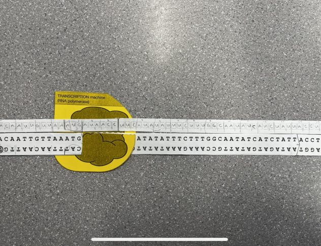
1. Describe the process of transcription
A section of DNA unzips and its bases are exposed. Complementary RNA nucleotides bond with bases on the exposed strand of DNA. Uracil is the complementary base that bonds with Adenine. Adjacent sugars and phosphates form bonds completing a strand of RNA that is complementary to the gene. This mRNA can now leave the nucleus for translation.
2. How did today’s activity do a good job of modeling the process of RNA transcription? In what ways was our model inaccurate?
The activity did a good job of modeling transcription by showing that the mRNA is formed by the complementary pairing of one of the DNA strands. It also shows that the enzyme used in this process is RNA polymerase. However, the model does not show that in reality, before the mRNA strand is formed, the DNA splits apart and after the mRNA strand is formed, it reattaches to itself. With our model, it looks as if the polymerase goes through the two strands of DNA as they are attached to each other, creating one strand of mRNA on one of the sides, which is inaccurate and could be misleading.
Translation:

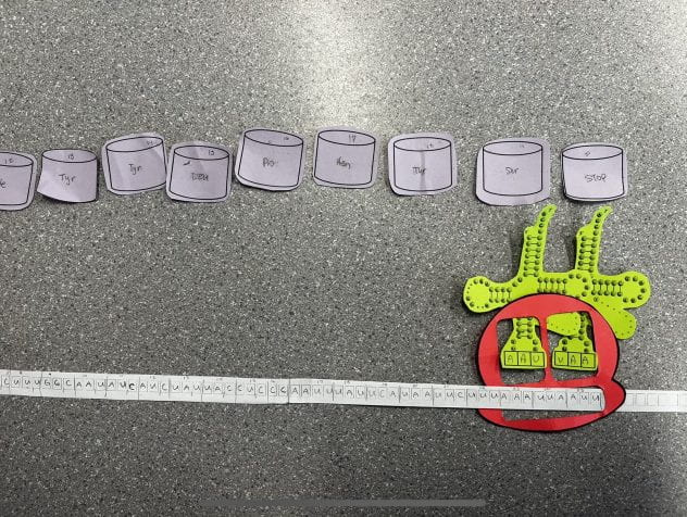
1. Describe the process of translation: initiation, elongation, and termination.
The first step in translation is initiation. Here, the mRNA with a START codon starts the translation process once attached to the ribosome. Then, the tRNA with a complementary anticodon to the mRNA codon holds an amino acid which will form a peptide chain in elongation. The ribosome continues to move over one codon at a time along the mRNA, attaching new tRNAs to the codon. More specifically, there is a P and A site to the ribosome. The initial tRNA enters the A side. Then, the next tRNA can enter the A site when the initial tRNA shifts to the P site. This repeats until a polypeptide chain forms. In termination, there is a STOP codon that is read on the mRNA. These do not code for any amino acids, rather there is a release factor that binds to the STOP codon on the A site. The completed protein is released from the ribosome and is sent for further processing.
2. How did today’s activity do a good job of modeling the process of translation? In what ways was our model inaccurate?
The model was great to show the polypeptide chain forming as the rRNA read the mRNA strand across the ribosome. We can use it to visualize how the chain forms and how the rRNA moves along the mRNA. From the model, we couldn’t show the tRNA moves from the A site to the P site because we were given only two tRNAs for the ribosome. In reality, there would be more tRNA in the cytoplasm that gets transferred in and out of the ribosome according to their complementary anticodon–codon pairs.
3. How is mRNA different than DNA?
DNA is double-stranded, whereas mRNA is single-stranded. DNA has deoxyribose as its 5-carbon sugar, and mRNA has ribose. DNA has thymine as one of the four nitrogenous bases, and mRNA has uracil in its place. DNA performs all of its functions and stays in the nucleus, whereas mRNA is small enough to travel from the nucleolus to the cytoplasm. DNA carries genetic information; mRNA determines the amino acid sequence of a protein. DNA replicates with the help of DNA polymerase, and mRNA is created by transcription with the help of RNA polymerase.