Explain the structure of DNA:
Deoxyribonucleic acid(DNA) is a macromolecule(nucleic acid) that are made up of monomers known as nucleotides. Each nucleotide is composed of a 5-carbon sugar, a phosphate group, and a nitrogen base. Each strand is composed of alternating molecules of deoxyribose and phosphate with nitrogen base attaching to each deoxyribose unit. There are two types of nitrogen bases, a purine (double ringed structure) and a pyrimidine (single ringed structure). The purines in DNA are Adenine (represented in yellow beads) and Guanine (represented in purple beads). My group had forgotten to add a second bead to these structures to represent the double ringed structure, but we recognize this structure and tried to represent it through their colours. The pyrimidines in DNA are Thymine (represented in blue beads) and Cytosine (represented in green beads).
The DNA strand consists of a sequence of nucleotides linked together to form a double helix that can also be described to appear as a long, twisted ladder. (This shape occurs as the final step of/in the DNA structure and is due to the forces caused by the bonds.) These strands run antiparallel to each other, which allows for complimentary base pairing through hydrogen bonding between nitrogen bases. Being antiparallel indicates that strands run in opposite directions but relay the same message. This means that one strand will start with the sugar while the other strand will start with the phosphate group. Complimentary base pairing describes how a purine is always paired with a pyrimidine. Adenine (represented in yellow beads) bonds with Thymine (represented in blue beads) and Guanine (represented in purple beads) binds with Cytosine (represented in green beads). The sequence of these bases are directly connected to the way our heredity information is coded in the genetic code in DNA & RNA.
See images below for a model of DNA structure:
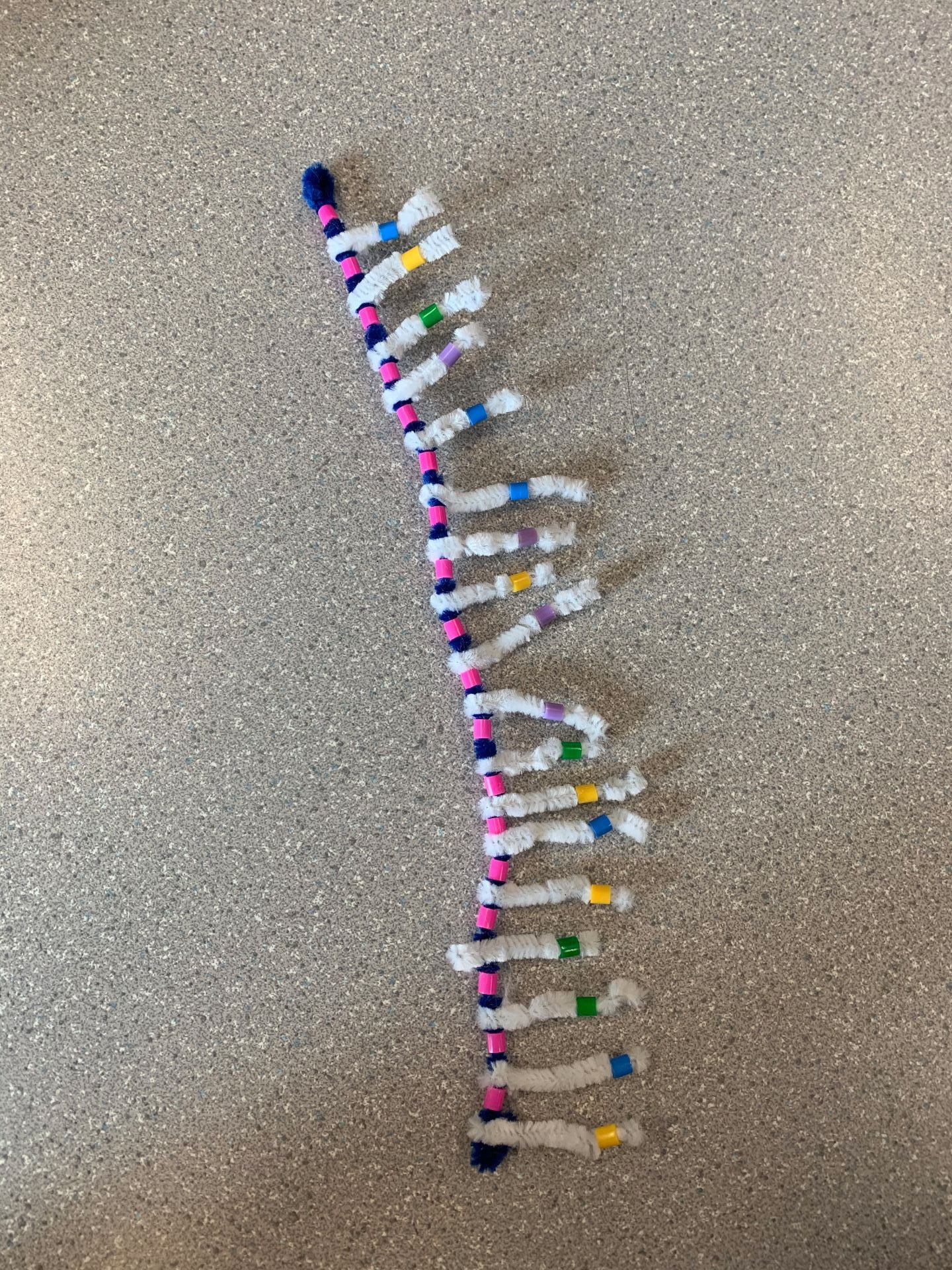
(This is an image of a single nucleotide. The blue pipe cleaner represents the sugar and the pink beads represent the phosphate groups which together make up the backbone of DNA. The white pipe cleaners represent the hydrogen bonds that will be made between nitrogen bases of two strands)
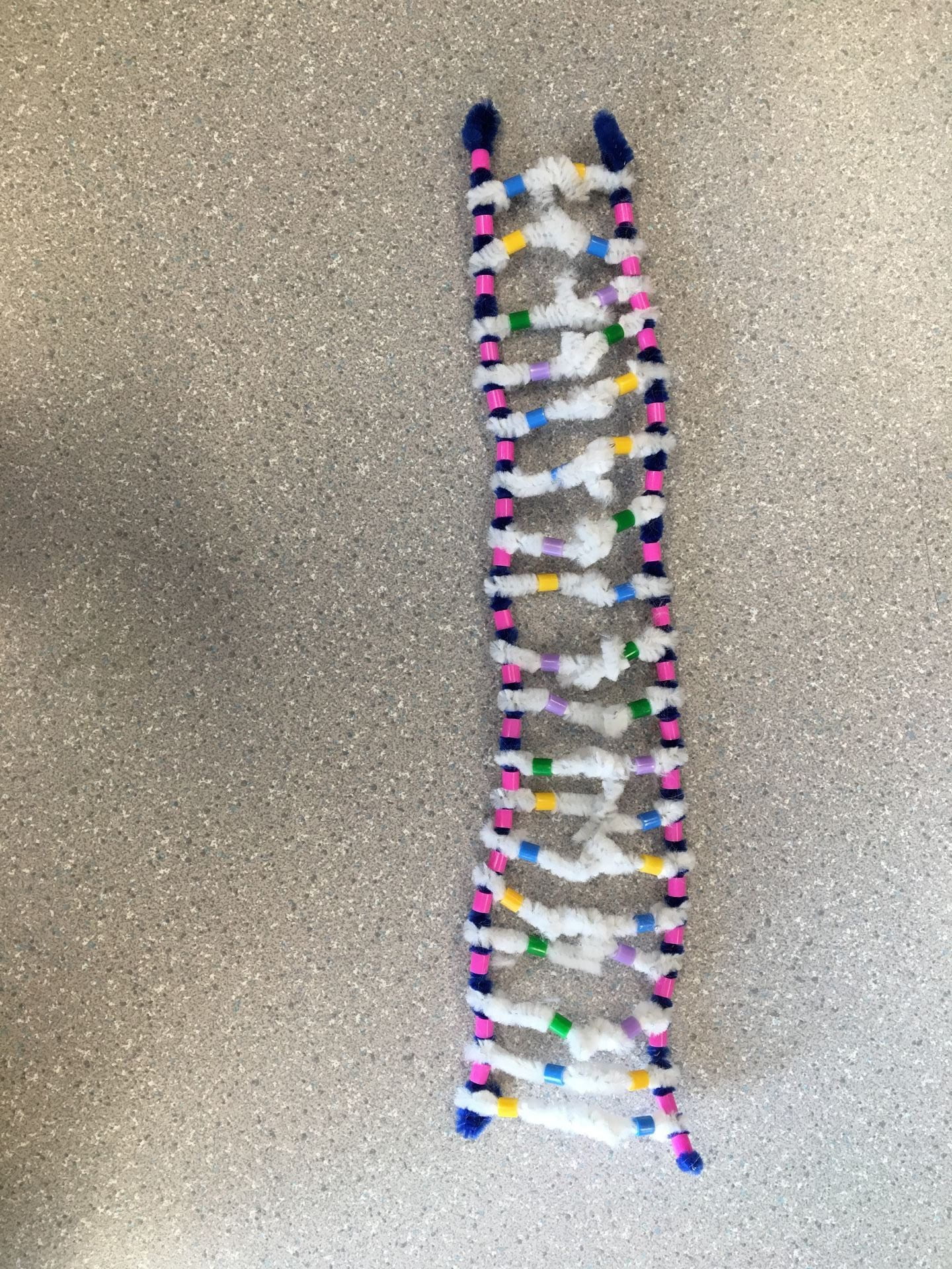
(In this image, we can see two nucleotides coming together to form complimentary base pairing between nitrogen bases. The white pipe cleaners represent the hydrogen bonds between the bases on opposing strands. Note how the strands are antiparallel; the strand on the left starts with a phosphate group while the strand on the right starts with the sugar.)

(This image represents the double helix shape that a DNA molecules makes.)
How does this activity help model the structure of DNA? What changes could we make to improve the accuracy of this model?
This activity provided a visual guide to the DNA structure process. We can easily see the phosphate groups, deoxyribose sugars, and nitrogen bonds through our usage of different coloured beads and pipe-cleaners. This model also demonstrated the complimentary base pairing process with hydrogen bonding with the two antiparallel strands. Overall, it really allowed me to grasp onto this concept with a high level of understanding.
As I mentioned before, my group had forgotten to add an extra bead for the Adenine and Guanine. We could’ve added this to indicate how these nitrogen bases are double-ringed. Some other changes could’ve been with ensuring that the sizing and proportions of the materials match. The model didn’t perfectly illustrate how a DNA molecule should appear as for example, the white pipe cleaners were not all exactly the same size and the spaces in between the sugar, phosphate, and nitrogen bases were not consistent throughout the entire strand. To improve this, we could ensure that all details are added and to try harder to equally space and place all materials. As well, DNA is normally extremely long so the size of the model we created is not and accurate representation of the size of a DNA molecule, however it would be almost impossible to create a physical model of a DNA molecule being its proper length.
DNA REPLICATION:
When does it occur?
DNA replication happens in all living organisms, it allows for genetic information to be passed down to be inherited. DNA replication is the process by which a DNA strand duplicates itself. Before a cell can divide, all the DNA must be duplicated meaning that DNA replication occurs prior to cell division, during mitosis. It is a semi-conservative process where each new DNA molecule contains 1 backbone from the original DNA strand and one from the new strand thus leaving us more DNA molecules than we started with.
Sequence of Events in DNA Replication:
- UNWINDING & UNZIPPING – the DNA double helix strand unwinds, and the two strands of DNA separate. This causes the hydrogen bonds between the nitrogen bases to break as well. DNA Helicase (represented by the green play dough) is the enzyme involved in this process. It unwinds the double helix and then works to break the hydrogen bonds.
- COMPLIMENTARY BASE PAIRING – new nucleotides move in to pair up with bases of each original strand of DNA. These nucleotides are free-floating around within the nucleoplasm. DNA Polymerase (represented by red play dough) is the enzyme used in this process. It connects the original strand to the new strand using complimentary base pairing, altogether creating an exact replica of the full original DNA molecule. –> (strands are still antiparallel)
- JOINING OF ADJACENT NUCLEOTIDES – sugar-phosphate bonds form between adjacent nucleotides of the new strand to complete the molecule. DNA Ligase (represented by blue play dough) is the enzyme responsible for attaching the nucleotides of the forming backbones to one another. It works to repair irregularities or breaks in the backbone of double-stranded molecules.
Leading vs. Lagging strands
On the leading strand, DNA synthesis occurs continuously. On the lagging strand, DNA synthesis restarts many times as the helix unwinds. This causes many short fragments called ‘Okazaki fragments’. The enzyme DNA ligase works to glue and join these fragments.

(This images represents the unwinding & unzipping stage/step in DNA replication. The green play dough represents DNA Helicase which works to unwind the double helix and break the hydrogen bonds.)
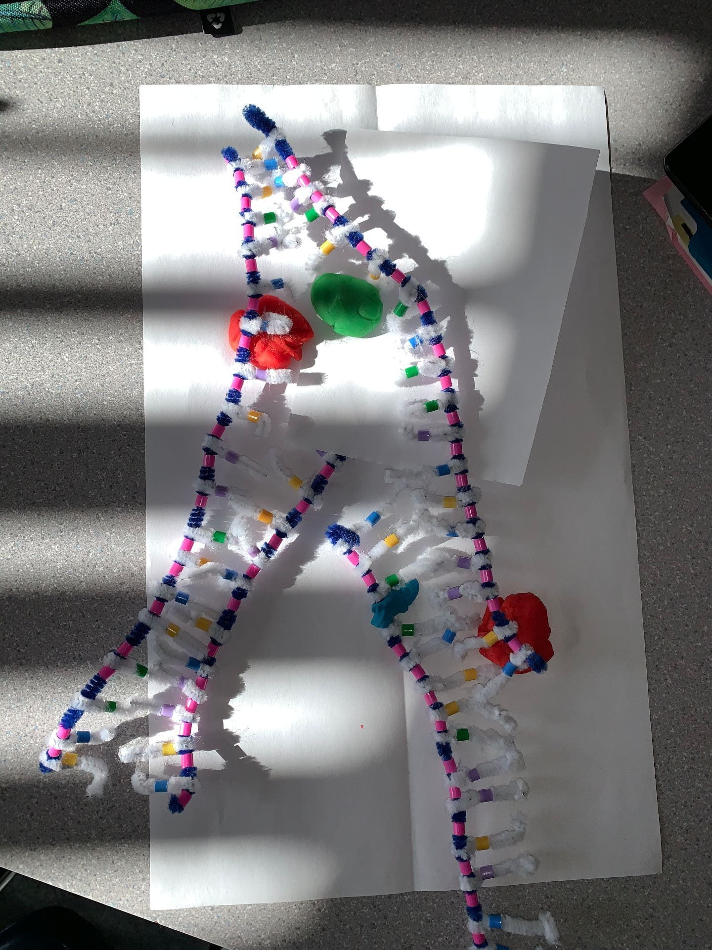
(This image is a combination of the complimentary base pairing and joining of adjacent nucleotides stages/steps in DNA replication. The green play dough is the DNA Helicase. The red play dough is the DNA Polymerase which is responsible for connecting the original strand to the new strand using complimentary base pairing, altogether creating an exact replica of the full original DNA molecule. The small bit of blue play dough is the DNA Ligase which works to repair irregularities or breaks in the Okazaki fragment in the lagging strand.)

(In this image, DNA replication is complete and we now have two full DNA molecules with both one strand from the original DNA molecule and one new strand.)
What Did You do to Model the Complimentary Base Pairing and Joining of Adjacent Nucleotide Steps of DNA Replication. In What Ways was This Activity Well Suited to Showing This Process? In What Ways Was it Inaccurate?
With this activity, I was able to model complimentary base pairing through the different colour beads and white pipe cleaners which acted as hydrogen bonds. The Adenine (yellow) always bonded with Thymine (blue) and the Guanine (purple) always bonded with Cytosine (green). We however weren’t able to illustrate the covalent bonds between nucleotides. The DNA Helicase, Polymerase and Ligase were all demonstrated using play dough which made it very easy to comprehend. My group had a hard time positioning everything correctly in our pictures of our models due to a small workspace and objects not staying still, which unfortunately allowed for some potential small spaces of discrepancy/inconsistency in our models.
This activity really gave me the opportunity to see exactly how the process of DNA replication works. This allowed me to understand the process better than if I were to simply hear/listen to the information. It also gave me an understanding of the different enzymes used in DNA replication and their roles. Also having to physically break bonds apart and add in different enzymes using play dough allowed me to grasp the concept much easier.
Altogether, while the models aren’t an exact replica of the DNA inside my body and while this activities had a few inaccuracies, I still found that it was very helpful.