In Biology 12, my class was assigned the job of creating a strand of DNA out of pipe cleaners beads. DNA is a molecule containing our genetic code. It is composed of a chain of nucleotides, monomers containing a 5-sugar backbone, a nitrogen containing base, and a phosphate. A DNA molecule is made up of 2 strands of chained nucleotides that is twisted into a double helix. Each strand contains a backbone formed by bonded sugar phosphate portions of adjacent nucleotides. Each nucleotide containes one of 4 types of nitrogen bases; are split into Pyrimidines (single ringed bases) and Purines (double ringed bases). Adenine and Guanine are Purines; Thymine and Cytosine are Pyrimidines. Two strands of DNA (which are anti-parallel) are hydrogen bonded together by the nitrogen bases of a nucleotide which connects the two strands together. Adenine specifically bonds with Thymine while Guanine bonds with Cytosine (Thymine and Adenine are double bonded; Guanine and Cytosine are triple bonded). A DNA molecule is around 85 nucleotides in length, therefor the order of the nitrogen bases (genetic code) determines which specific proteins are made and which functions are to be done.
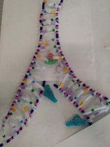
The model below is my groups representation of a 18 nucleotide DNA molecule with the materials listed in the beginning. The blue pipe cleaners represents the sugar backbone of the DNA molecule, pink beads represent the phosphates connected along the sugar backbone, the colored beads represent the nucleotide bases (Purple is Guanine, Green is Cytosine, Yellow is Adenine, and Blue is Thymine; the number of beads represents the number of rings for each nitrogen base – 2 for Pyrimidines and 1 for Purines), and the white pipe cleaners represent the hydrogen bonds between the base pairs. This activity is a good representation of what a DNA stand looks like. My group and I were able to show the sugar phosphate backbone using the blue pipe cleaner and pink bead as well as the different nitrogen bases with different colored beads connected by white pipe cleaners representing the hydrogen bond. The only difference I would make to further enhance the model is possibly using another colored bead onthe hydrogen bond pipe cleaner to represent the number of bonds between nucleotides; 2 for A and T and 3 for G and C.

 Next our group was asked to show DNA replication. DNA replication is a process where a cell will make a copy of its DNA so that during Mitosis (Cell Division) each cell has the genetic information to continue dividing and carry out cell functions. There are three possible ways of DNA replication, the correct way being semi-conservative replication (each daughter molecule has one parent strand and one daughter strand; other two possibilities are conservative and dispersive replication). During replication, an enzyme called DNA Helicase unwinds and unzips a DNA molecule splitting the two strands apart. New nucleotides are created as the molecule is unzipping and are bonded by DNA Polymerase on to each strand. The nucleotides are being built in the direction from 3 Prime towards 5 Prime in reference to the parent strand however, the way new nucleotides are being connected differs between the two strands. During DNA replication, one strand is “leading” and the other is “lagging;” the leading strand has nucleotides built in the direction of the replication fork, the lagging strand is being built in the direction away from the replication fork in sections called Okazaki fragments that are glued together by an enzyme named DNA Ligase. These processes continue until the original parent molecule completely unzips into two parent strands and new daughter strands are being built onto the parent strands as the molecule unzips.
 The model to the leftis representing the process of DNA replication. The materials that represent the DNA molecule are the same as that of the image above. The Blue candy however represents the DNA Polymerase which bonds the new nucleotides onto the parent strands while the peach candy represents DNA Helicase that unwinds and unzips the DNA molecule. The model on the right shows the result of DNA replication: two new Daughter molecules that each contain one parent and one daughter strand. Modelling DNA replication was very difficult because the I feel these images do not represent the process effectively. Although we were able to show the unzipping, the lagging strand is hard to identify.

To so however, as you can see, to represent the anti-parallel strands, one strand has a phosphate on top (representing 3 prime) and the other strand has a phosphate on the bottom with no phosphate on the top (representing 5 prime). Even though we did this, it is hard to see in the picture to the left. Another problem was not that the model was inaccurate however we were unable to show how the lagging strand is built upon. We know the lagging strand has nucleotides that are being built away from the replication fork in okazaki fragments and in the model we did not show this. Overall, I believe both models of the DNA structure and DNA replication do a good job in showing how the molecule looks like at a macroscopic level.
