 For this activity, my class was asked to model out Protein Synthesis: Transcription and Translation. Similarly to the DNA Structure and Replication assignment, we modeled the processes using pipe cleaners and beads. Firstly in Protein synthesis, DNA undergoes Transcription: DNA replicates one of its genes into forming Messenger Ribonucleic Acid (mRNA), a molecule similar to DNA, which is made up of a chain of nucleotides. Each nucleotide, like DNA, contains a 5-carbon sugar (Ribose), a phosphate, and a nitrogen base. The RNA backbone is composed of Ribose sugars and phosphates. There are 4 different nitrogen bases that are divided into two groups Purines and Pyrimidines. Guanine and Adenine are Purines while Cytosine and Uracil are Pyrimidines. In RNA, the base: Uracil, replaces Thymine as the complimentary base to Adenine, while Guanine is complimentary base pairs with Cytosine. There are two major differences between the two molecules. The first difference with RNA and DNA is that DNA is composed of two backbones that are Hydrogen bonded together while RNA is made up of only 1 backbone. The second difference between DNA and RNA is that DNA is composed of over 85 million nucleotide while RNA is composed of around 1000 nucleotides; RNA is a copy of 1 gene of DNA. The process of Transcription is very similar to DNA replication. A section of DNA unwinds and unzips, exposing 1 gene. Along 1 strand of DNA, complimentary bases are added on to form the nucleotide chain. Next, RNA Polymerase (an enzyme similar to DNA Polymerase) joins adjacent nucleotides together to form the RNA backbone. Finally, the RNA is released and the DNA reforms it’s double helix. Following Transcription, the RNA leaves the nucleus of the cell (where the genetic information: DNA is held) and moves into the Cytoplasm.

The picture to the left is our groups model of Transcription in the middle of the process. Similar to the previous activity, the DNA backbone is composed of Blue pipe cleaner and pink beads representing the 5 Carbon sugars and phosphates, the colored beads represent the nucleotide bases (Purple is Guanine, Green is Cytosine, Yellow is Thymine, and Blue is Adenine; the number of beads represents the number of rings for each nitrogen base – 2 for Pyrimidines and 1 for Purines), and the white pipe cleaners represent the hydrogen bonds between the base pairs.

The RNA strand is composed of a red pipe cleaner and pink bead backbone (5-Carbon sugar and phosphate), colored beads representing the individual nitrogen bases (the same as the DNA strand except the Yellow Adenine is replaced with a brown Uracil – 2 beads showing it is a pyrimidine). The peach candy represents the DNA Polymerase adding the new nucleotides together to form the RNA molecule. The picture on the right shows the end result of this process. As a result, 1 strand of RNA is made carrying the genetic information of 1 DNA gene to form the protein while the DNA molecule reforms itself and twists back into the double helix shape.
 The next step in Protein synthesis is Translation. When mRNA is created, it leaves the nucleus of the cell and enters the cytoplasm where Ribosomes are located. The Ribosomes’ job is to build a polypeptide chain of amino acids that forms a protein. It does this by reading the order to the nucleotides of the mRNA like a code and each codon (group of 3 nucleotides) contains the coding (based on order of nucleotides) for a specific amino acid that is added onto the polypeptide chain. In Translation, there are 3 major phases that take place.
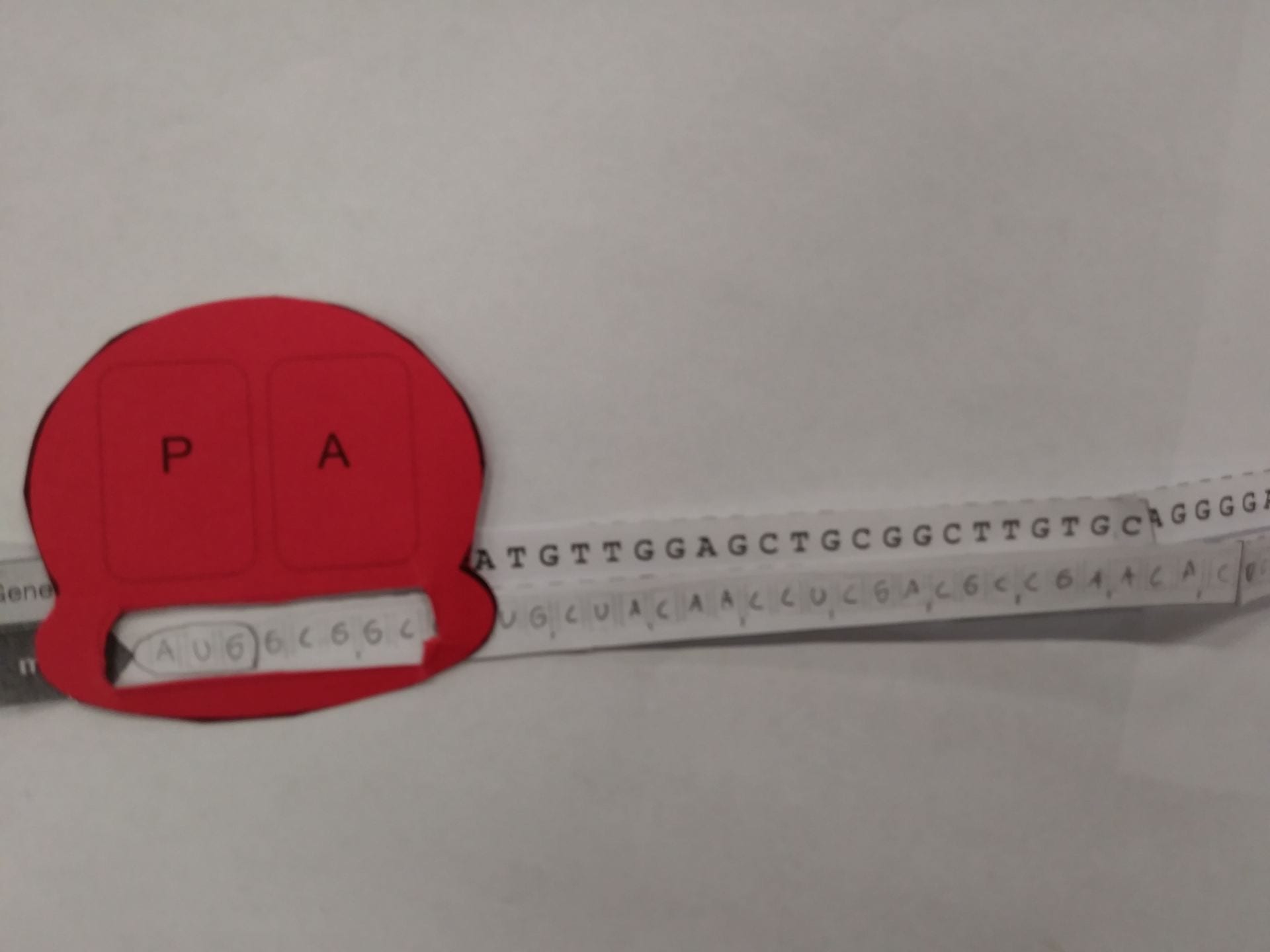
 The first phase in Translation is Initiation: The mRNA is held by the Ribosome where the code is read. The Ribosome starts by reading the Start Codon (AUG) in the P-site, where then another form of RNA: tRNA collects the amino acid that is paired with the anti-codon of the current codon being read.
 The second step is Elongation: The chain continues to be built. Now the A-site of the Ribosome reads the code of the mRNA, were the tRNA in the A-Site locates the amino acid that is coded for the codon and and then added on the the amino acid chain in the P-site.
 The final step is Termination, where the Ribosome reads a stop codon where it is instructed to stop the process, resulting in the Ribosome letting go of the mRNA and the tRNA to let go of the chain of amino acids.