
1a) plant cells under a microscope look very transparent. There’s not much to see since the membrane of the onion was very thin. We did however see scale-like patterns in the background that had hints of light purple.
The cell structures I see are chloroplasts, cell membrane, cytoplasm and the cell wall.
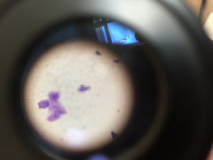
1b) Animal cells have tiny shards floating around everywhere. Some are attached together. The background is clear but has little grey dots everywhere.
The cell structures I see are cytoplasm, the nucleus and the cell membrane.
- In my opinion, the way I can mostly tell plant and animal cells apart is what’s behind the cell structures. I found that animal cells don’t have much of a background than plant cells. Plant cells have a scaly background and animal cells do not. Another way I can tell them apart is how they are formed. Plant cells have a square like shape and animal cells have a circular shape.
- We use methylene blue on an animal cell because for this occasion the cells were transparent meaning that id we put it under a microscope we wouldn’t see anything since its clear. By adding pigment to the cells, it allows us to clearly see all the different types of cells.
We do not use it on plant cells because plant cells normally have colour so we can automatically see the cell structures without adding colour.
- I learned more about microscopes and how to properly focus objects using dials. I learned that it is very hard to see and observe animal and plant cells when looking for cell structures. Finally, I learned that you must first dye animal cells blue to see all the cells.
Questions:
Can we put more items under the microscope next time?
Do we always need to use methylene blue on animal cells?
Can we observe cells that move in real time under the microscope?
How does methylene blue work?
Curious:
What happens when we put a piece of fur underneath the scope?
What will we see if we put a piece of dust, food and clothing underneath?