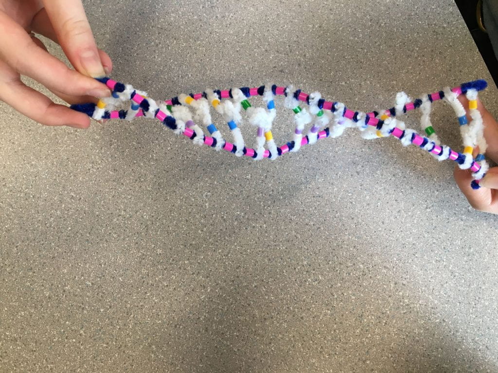
DNA and mRNA both play very important roles in our bodies but differ in some ways.
- DNA is double stranded and appears as a double helix shape, mRNA is only single stranded and appears as a single spiral.
- DNA is made up of deoxyribose sugar while mRNA is made up of ribose sugar.
- mRNA uses the nitrogen base Uracil instead of Thymine. It is still a pyramidine that hydrogen bonds with Adenine. (the rest of the nitrogen bases remain the same along with their complimentary base pairings)
- mRNA is much smaller than a DNA molecule as it travels in and out of the nucleus through a nucleus pore.
- DNA contains the genetic instructions of living things while mRNA is produced from DNA and carries DNA’s message to the ribosomes.
- DNA is created through DNA replication while mRNA is formed through a process called transcription
- mRNA is primarily found in the cytoplasm of cells while DNA is found mainly in the nucleus
Describe the process of transcription:
Transcription is the process by which the information in a strand of DNA is copied into a new molecule of messenger RNA (mRNA). This transcription process occurs in multiple steps, all of which occur due to the enzyme, RNA polymerase.
1. UNWINDING & UNZIPPING OF DNA – A specific section of the DNA molecule unwinds, exposing a set of bases (a gene). The hydrogen bonds between nitrogen bases in this specific section have been broken to allow for RNA polymerase to come and continue the transcription process.
(this is an image of the original DNA molecule in its double helix form. This is before the transcription process begins)
(In this image, the DNA molecule has unwinded and now looks like a flat ladder. Note that the unzipping step has not been included in this image)
2. COMPLIMENTARY BASE PAIRING WITH DNA – RNA polymerase reads the unwound DNA strand and builds the mRNA molecule, using complementary base pairs. This is only along one strand, the sense strand. This is the strand that carries the correct instructions for building the protein, unlike the nonsense strand which if transcribed would would not yield a protein. Complementary base pairs are made between nitrogen bases (these are hydrogen bonds). This is the same concept as in DNA molecules, however, since RNA (represented by a red pipe cleaner) does not contain thymine as its pyrimidine nitrogenous base, it has uracil (represented by brown beads) to pair with adenine instead (guanine and cytosine will still pair together). The RNA polymerase is also responsible for the forming of covalent bonds between adjacent nucleotides to build the RNA backbone.
(My group took this photo to represent the complimentary base pairing step in transcription. Our model does include some inaccuracies which will be explained in detailed in the following question. Note that Uracil is represented by brown beads and that the mRNA strand has been represented by a red pipe cleaner.)
(please refer to this image as an accurate representation of this DNA transcription step.)
3. SEPARATION FROM DNA – This is the last step in the DNA transcription process. The mRNA separates from the DNA after the entire gene has been transcribed. The DNA reforms its double helix shape by zipping and winding back together. The mRNA is now leaving the nucleus and starting the translation process. At this step, we should be left with a DNA molecule in a double helix shape and an mRNA molecule in its “spiral-esque” shape.
(This is image shows the reattached DNA molecule and the mRNA molecule. Note that both have not been positioned into their correct structural shape and should not be touching.)
How does this activity do a good job of modelling the process of RNA transcription? In what ways was our model inaccurate?
There was some confusion about the representation of the steps of transcription within my group which ultimately led to some inaccuracies in our visuals. We had the RNA and one of the DNA strands fully bonded together when we should’ve illustrated it in a way that had the DNA still attached at one end and unzipped at the other end allowing the mRNA to bond with the sense strand of DNA. We did however illustrate the RNA polymerase correctly for this model. Our model was also inaccurate in the way that the DNA molecule and the mRNA molecule are the same size. A DNA molecule is much larger than an mRNA molecule. In RNA transcription, only a portion of DNA is used to make an mRNA molecule. In the model, the entire DNA molecule is used. Despite the inaccuracies produced by my group, I still think that this activity was beneficial and really helped me to grasp the transcription concept by providing visual aids.
Describe the process of translation: initiation, elongation, and termination:
Translation occurs right after transcription. It is the process of of translating the sequence of a messenger RNA (mRNA) molecule to a sequence of amino acids during protein synthesis. Similarly to transcription, the translation process occurs in multiple steps…
*Things to understand before looking at translation!*
- In an mRNA, the instructions for building a polypeptide come in groups of three nucleotides called codons. We use these codons to figure out which amino acids are involved and in what order. We find the amino acid using a special “protein sheet” from our three nucleotides which are labelled based on the nitrogen base of the mRNA strand. There is always a start and stop codon.
1. INITIATION – Translation begins with this stage. mRNA is being help by the ribosome. The P site of the ribosome reads the AUG start Codon on the mRNA strand (the start codon is ALWAYS AUG (methionine). The tRNA will bring in the first amino acid and then it will continue this process (elongation) until a stop codon (UGA) is reached.
2. ELONGATION – In this step, the amino acid chain continues to grow. The A-site reads the next mRNA codon and bring in the matching tRNA. The amino acid chain from the tRNA in the P-site is transferred to the tRNA in the A-site. This continues to create a polypeptide chain.
(In this image we can see the start codon and its amino acid in the P-site plus the second mRNA codon in the A-site. Note that my group did not include the following step where the amino acid chain from the tRNA in the P-site is transferred to the tRNA in the A-site.)
3. TERMINATION – This is the last stage of translation and allows for the polypeptide chain to hopefully become a fully functioning protein. In order for this to occur, we must reach one of the three stop codons. There are no more amino acids onward. The ribosome lets go of the mRNA and the tRNA lets go of the polypeptide.
(this image shows my group’s finished amino acid chain. Note that the blue pieces of paper should not be side by side and should actually be stacked one above the other on top of the green pieces of paper from the image before.)
How does today’s activity do a good job of modelling the process of translation? In what ways was our model inaccurate?
My group experienced a little bit of confusion with how many pictures we needed to take. So, although we completed the whole process of translation and understand the steps, we were not able to show through our images each detailed step because we didn’t end up taking certain photos. There was also some issues with spacing which forced some of our papers to not line up properly to create a slightly altered/inaccurate representation of that specific step. The importance of the start and stop codons was highlighted which really helped me to grasp their importance in this translation process. Altogether, it is simply just hard to fully accurately show how this process occurs with pieces of paper but we tried our best and I think I still benefited from this activity. It demonstrated a pretty good image of what translation really was even with a couple of small details missing.


