Explain the structure of DNA – use the terms nucleotides, antiparallel strands, and complimentary base pairing.
DNA is a large polymer that is constructed of nucleotide monomers. DNA is built of phosphate, a nitrogen base (pyramidines and purines), and a pentose sugar (deoxyribose). DNA (deoxyribonucleic acid) consists of two backbones that are made of repeating sugar-phosphate units. DNA is twisted into a double helix shape. The backbones of DNA are antiparallel which means that one of the strands goes from 3’-5’ and the other goes from 5’-3’. The 3’ indicates that the side has a hydroxyl group and the 5’ represents the side with the phosphate group. The strand that starts with 3’ is the leading strand and the strand that starts with 5’ is the lagging strand. Since DNA is read from 3’-5’ the strands are read in the opposite directions. The nucleotide bases on the backbones extend inward and they also form H-bonds between their complimentary base which causes the rung structure of DNA. The nitrogen bases used in DNA are from two different groups purines (Adenine and Guanine) and pyramidines (Thymine and Cytosine). Purines have a double ring base and pyramidines have a single ring base. The four nucleotide bases of DNA are Adenine, Guanine, Cytosine, and Thymine Adenine has to bond with Thymine, its complimentary partner, and Cytosine has to bond with Guanine, its complimentary partner. The complimentary base pairing is required so that the message on the strands can be read.
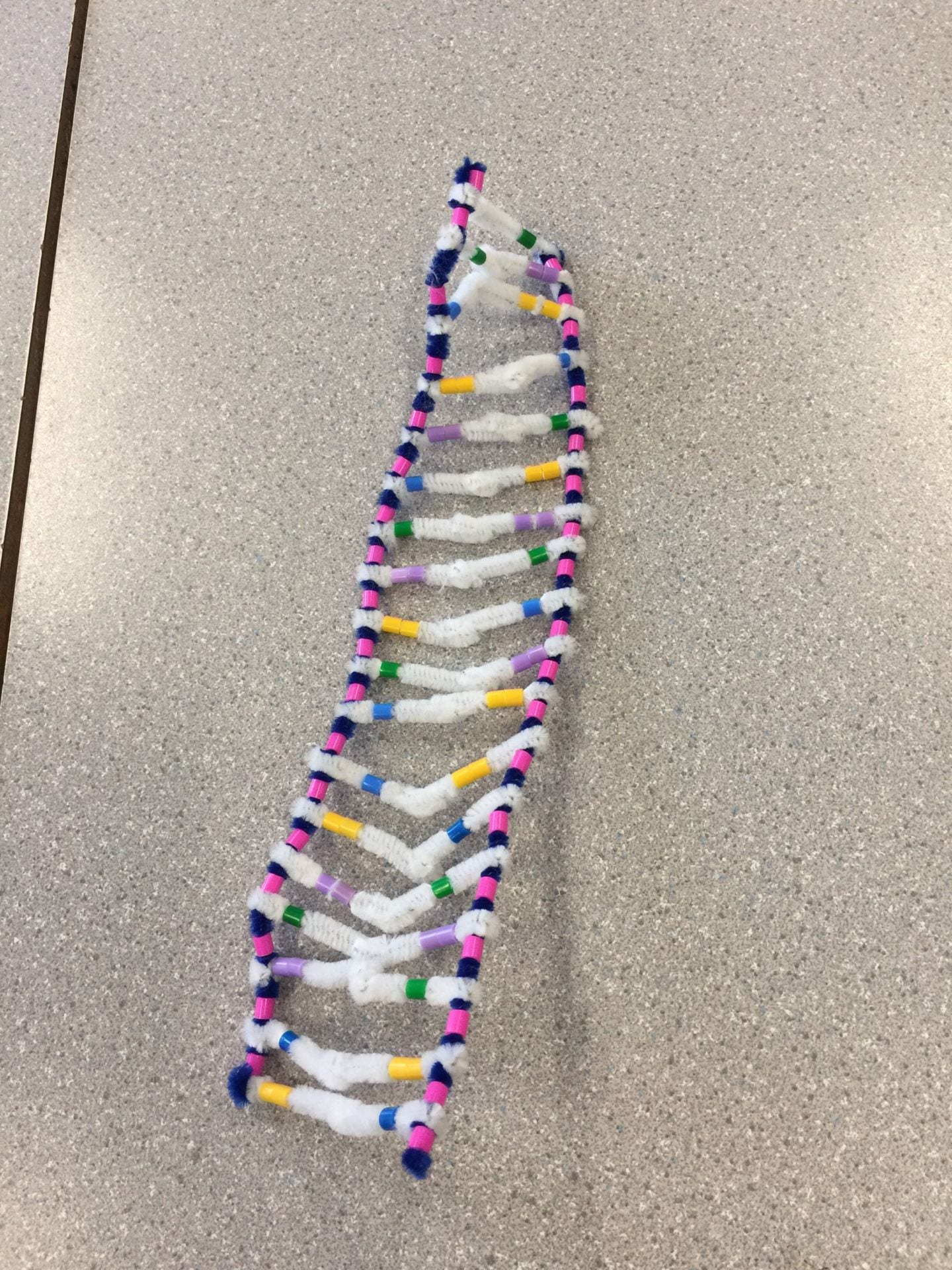
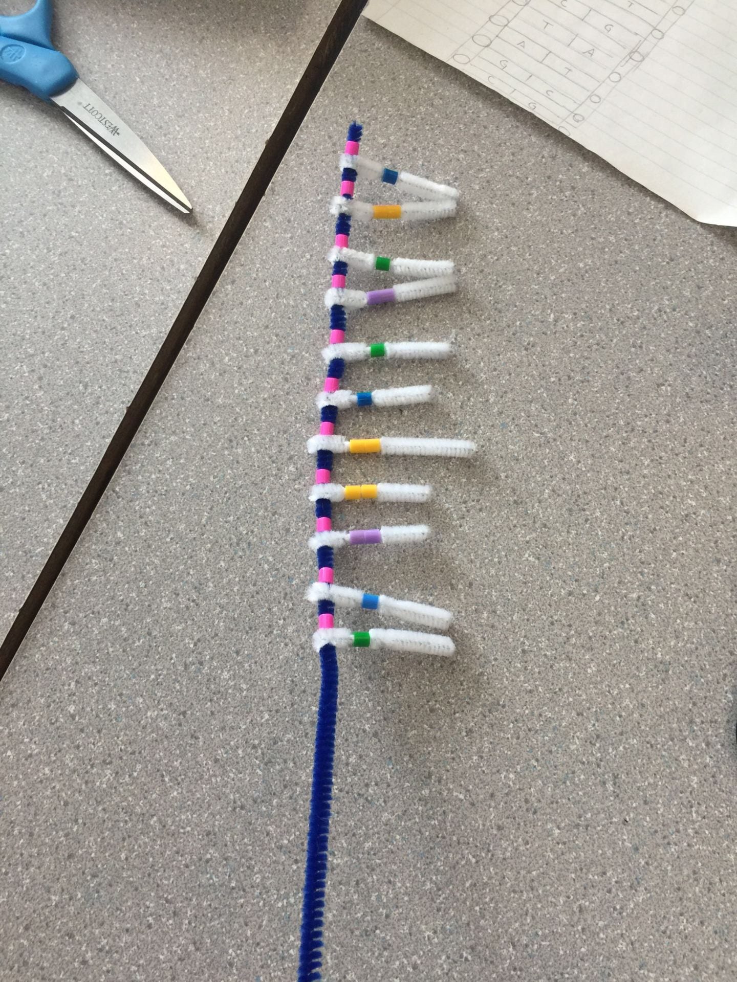
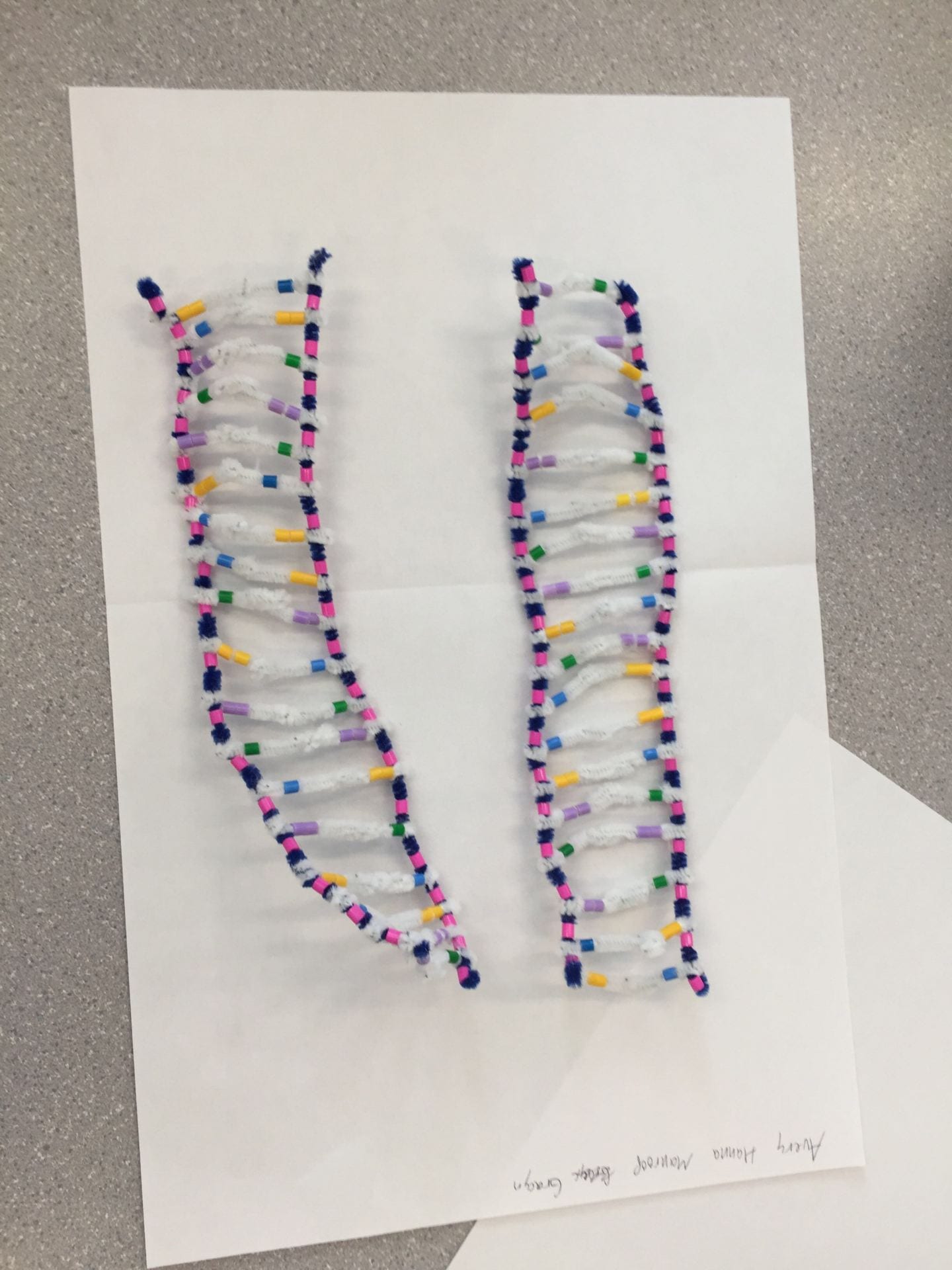
In the picture below is our DNA model. The blue pipecleaner represents the backbone and the pink beads represent the phosphate to show the sugar-phosphate backbone. The antiparallel structure of the DNA strands is shown in the model by the pink beads. At the top of the left backbone there is a pink bead (phosphate) but at the top of the right backbone there is no pink bead, similarly at the bottom of the left backbone there is no pink bead, but on the bottom of right backbone there is a pink bead. You can also see that the green beads (cytosine) are always paired up with the purple beads (guanine) and the yellow beads (adenine) are consistently paired with the blue beads (thymine), this represents the element of complementary base pairing in DNA. The beads are connected by a white pipecleaner which represents the H-bonds that are formed when they are paired together. The picture on the left clearly shows the rungs formed by the H-bonds and the picture on the right shows the double helix shape of DNA.

How does this activity help model the structure of DNA? What changes could we make to improve the accuracy of this model? Be detailed and constructive.
This activity was a great way to visualize the actual structure of DNA because you could clearly see the antiparallel strands, H-bonds formed by the bases, the different bases (double and single), and sugar phosphate backbones. One of the things that could be better indicated in this activity could be the 3’ and 5’ ends of the backbones because you couldn’t clearly see which side was which. It was also hard to keep our phosphate beads from moving in between the H-bonds of the nucleotides, which made it a little difficult to show the antiparallel strands.
PART II:
When does DNA replication occur?
DNA replication occurs in the nucleus of a cell. DNA replication takes place during interphase right before cell division. In order for cells to divide they have to double their genetic information, which is why DNA replication is a vital step for cell division. DNA replicates itself trillions of times in a lifetime. Each replicated DNA molecule has a strand from the original DNA molecule (parent) and a new strand (daughter).
Name and describe the 3 steps involved in DNA replication. Why does the process occur differently on the “leading” and lagging” strands?
Unwinding:
The first step of DNA replication is unwinding. The two backbones unzip which means that the H-bonds between the nucleotides break. The DNA Helicase enzyme causes the unwinding of the backbones. This process splits the leading strand and the lagging apart. These two strands are then going to be used as templates to make the daughter strands.
Complimentary Base Pairing:
The next stage of DNA replication is complimentary base pairing. Now that the backbones are split the daughter strand can now get the correct nucleotides in place to replicate the parent strand. Cytosine still bonds with guanine and adenine bonds with thymine, this makes the antiparallel strands. This process is caused by the enzyme DNA polymerase which causes the bases on the parent strand to have their complimentary partner. DNA polymerase can only copy strands in the 5’-3’ direction, this causes the lagging strand to be copied in a number of sections and not straight down. This also causes the lagging strand to take a longer time to replicate its DNA.
Joining:
The last step of DNA replication is joining. In this step the complimentary bases form H-bonds to join together and the sugar and phosphates form covalent bonds to join together. On the leading strand, this process is continuous as the DNA molecule unzips. Whereas on the lagging strand, there are fragments of nucleotides which require the enzyme DNA ligase to join these fragments together.
The processes are different depending on the type of strand because a lagging strand has to go through extra steps. Since DNA polymerase cannot copy the strands in the 3’-5’ direction there are segments of the lagging strand that are missing. DNA polymerase eventually goes back to these segments and pairs the nucleotides with their proper complimentary partner.
The picture below shows the process of replication occurring. The watermelon represents the enzyme DNA helicase. The watermelon continues to move up the DNA molecule to unzip the backbones. The blue bigfoot represents DNA polymerase which is why it is on both the lagging and leading strands. The red bigfoot is only on the strand on the right because it is the lagging strand. As you can see in the picture, the strand on the right has fragments of nucleotides missing.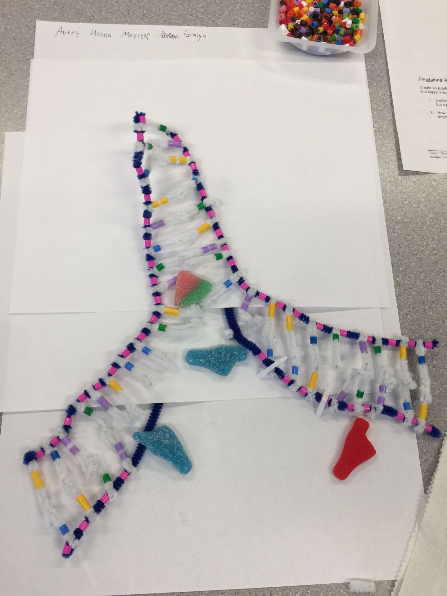
The result of DNA replication is two DNA molecules that are identical to each other.
The model today wasn’t a great fit for the process we were exploring. What did you do to model the complimentary base pairing and joining of adjacent nucleotides steps of DNA replication? In what ways was this activity well suited to showing this process? In what ways was it inaccurate?
In order to show complimentary base pairing, we placed the blue bigfoot in the areas where the bases had just been paired up. I think that we could have done a better job by clearly showing that the bases had not bonded together yet and were still waiting to go through the joining process. We showed the joining of adjacent nucleotides by placing the red bigfoot in the area of the missing segments of the lagging strand. I think that this activity helped me gain a stronger understanding of the general process of DNA replication because we had to go through each of the steps. I think that this activity didn’t go very in depth with the process of polymerase because we didn’t address the use of RNA primase. We could’ve shown the process of ligase better by having more fragments on the lagging strand.