Explain the Structure of DNA:
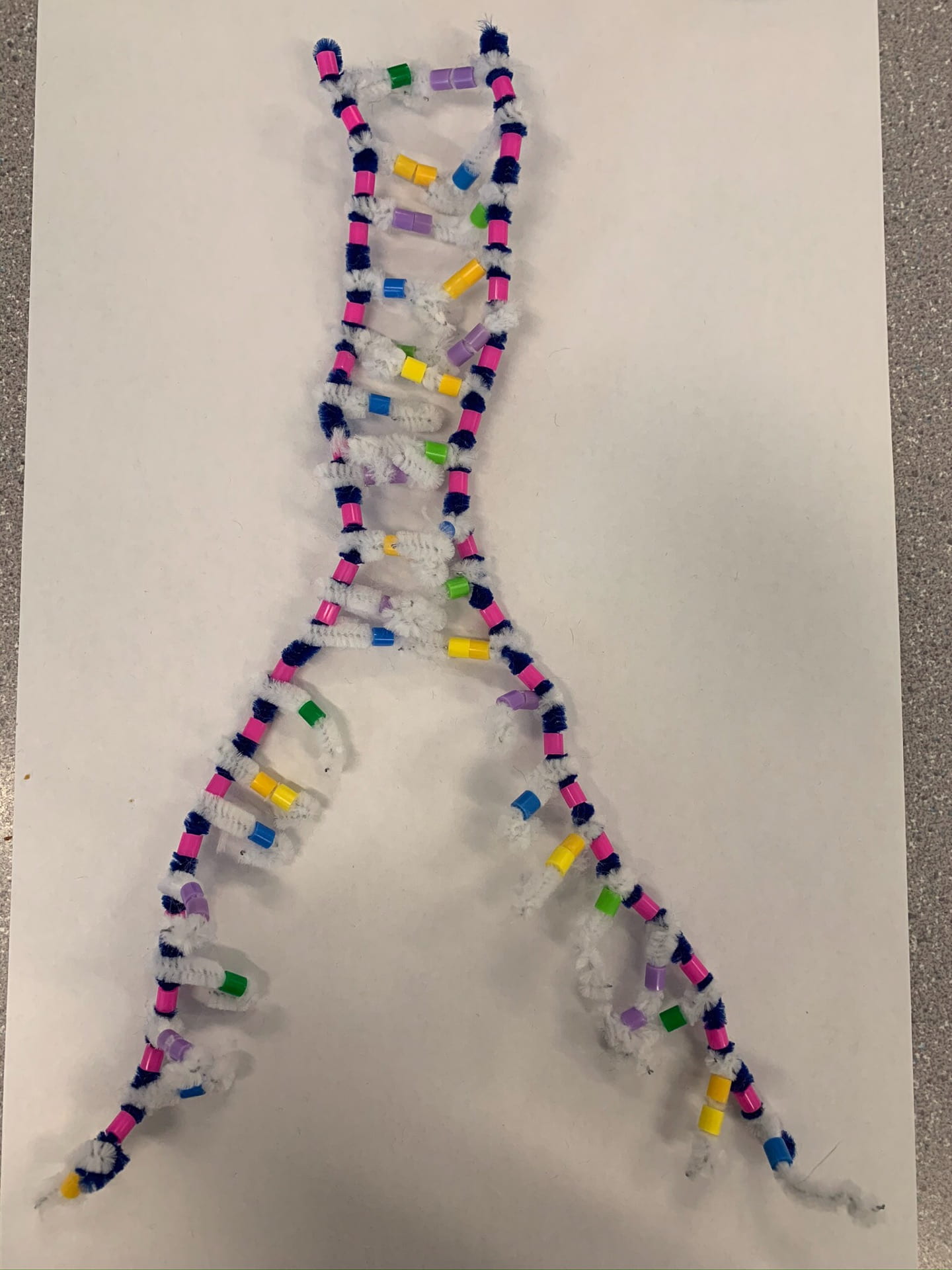
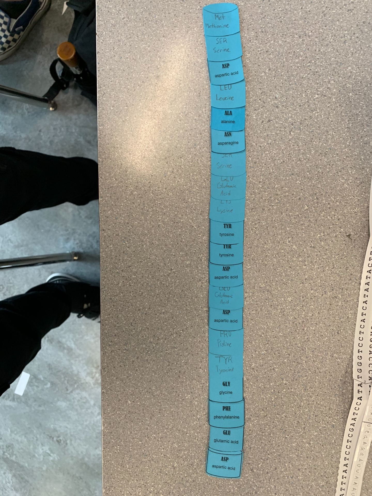
DNA (Deoxyribonucleic acid) is a nucleic acid made of monomers of nucleotides. Nucleotides include a Phosphate group, a 5-carbon sugar, and a nitrogenous base. There are two types of these nitrogenous base’s, purines and pyrimidines. Purines are a double ringed structure (2 beads), while pyrimidines are single ringed (single bead). The purines in DNA are adenine and guanine and the pyrimidines are cytosine and thymine. DNA has two strands, which are antiparallel meaning they run in opposite directions. The head of one strand is always at the tail of the other strand (one strand is 5’ to 3’ the other is 3’ to 5’). The pink bead in the picture below symbolizes the phosphate. On the left strand the pink bead (phosphate) is the head (5’) but the strand on the right has the 5’ at the bottom.
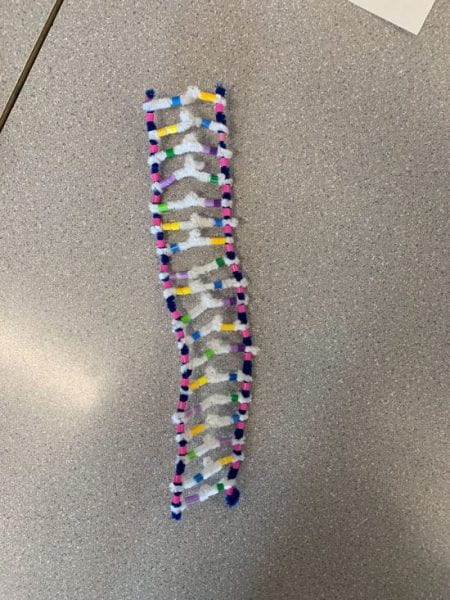
The white pipe cleaners show bonding between the two strands, through Hydrogen bonding. This is also when the town nitrogenous bases are paired together with complimentary base pairing. This means that cytosine and guanine in DNA are bonded together with 3 hydrogen bonds and adenine and thymine are paired together with 2 hydrogen bonds in between. As shown above there are specific rules for complimentary base pairing. Yellow and blue (thymine and adenine) and purple and green (cytosine and guanine). After these two strands are bonded together through complimentary base pairing the monomer nucleotides. The final step of the structure occurs, the strands twist together through forces between the bonds, this is called double helix. It is visually displayed below.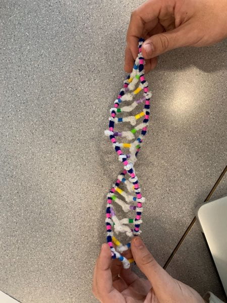
How does this activity help model the structure of DNA? What changes could we make to improve the accuracy of this model?
It gives you a prime visual of what DNA looks like. For me personally having the white pipe cleaner showing the H-bond between the strands is great. Also the beads show purines with 2 and pyrimidines with 1. Then having the blue pipe cleaner resemble the 2 Strands of nucleotides backbones and pink beads as the phosphate. It gives me a great visual of all the pieces of DNA. However it would be cool to somehow have a glimpse of how long they are, for this model only shows about 20 nucleotides long, when DNA can be up to 85 million long. To improve accuracy maybe we could show the 5-carbon sugar more accurately by having another bead for it, and then putting the phosphate to the side of it somehow because technically they aren’t on top of each other as this model shows.
When does DNA Replication Occur?
DNA replication happens in all living organisms. This process happens before cell division, and passes down genetic information to be inherited. It’s called a semi-conservative process where the replicated strands keeps an original strand and a replicated strand to create a brand new DMA in a double helix.
Name and Describe the 3 Steps Involved in DNA Replication. Why Does the Process Occur Differently on the “Leading” and “Lagging” Strands?
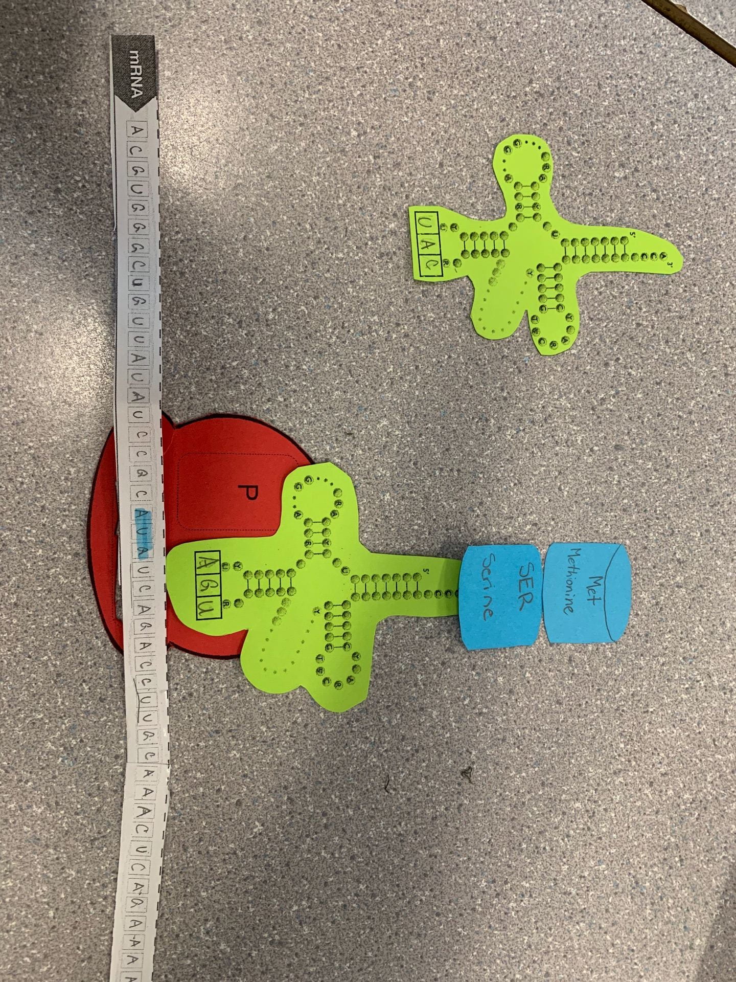
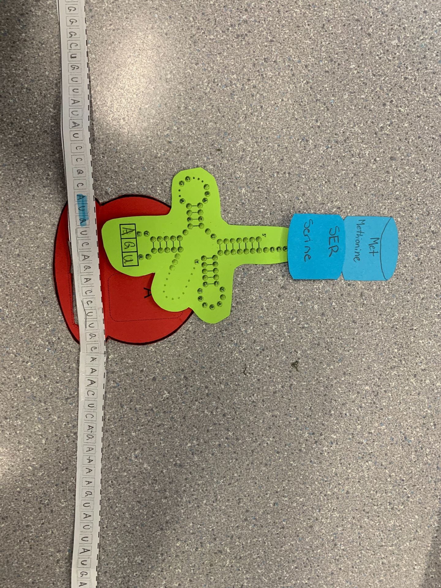
Unwinding and unzipping, something called DNA Helicase unwinds the double helix so it’s like a straight ladder. Them it unzips by breaking through the H-bonds, as The green enzyme shows below.
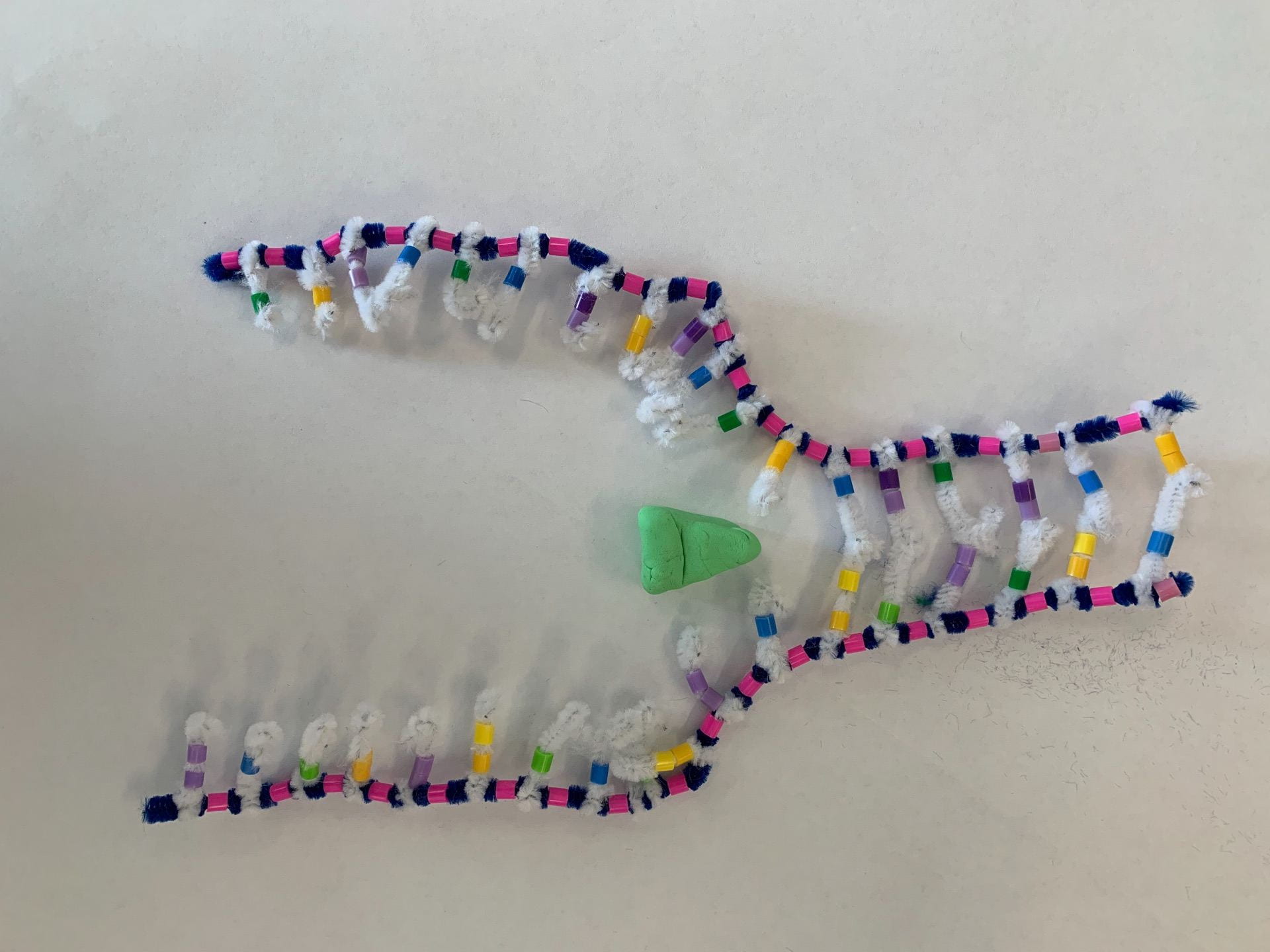
Complimentary base pairing, so after the strands are unzipped DNA Polymerase uses the free-floating nucleotides in the nucleoplasm to pair them to the unzipped strands. DNA Polymerase can only build from 5′ to 3’, so it cannot go 3’ to 5’. This first strand can work without interruption and is know as the leading strand. Then the other strand has to work backwards because it has to go 5’ to 3’ resulting in Okazaki fragments. This strand is called the lagging strand.
Adjacent nucleotides joining, the individual nucleotides that we’re bonded in the process before are now bonded beside each other with a sugar phosphate backbone. The enzyme DNA Ligase is responsible for this. And then finally it’s capable of winding into a double helix strand, creating a complete DNA strand.


What Did You do to Model the Complimentary Base Pairing and Joining of Adjacent Nucleotide Steps of DNA Replication. In What Ways was This Activity Well Suited to Showing This Process? In What Ways Was it Inaccurate?
In this this activity we showed the phosphate bonds and the nucleotides joining at the same time, this is inaccurate. However, it was a good example of the unzipping and the creation of the template strands.