April
17
April
11
Piecewise Differentiable Function with Tangent Line Surfer Desmos Project – Calculus 12


January
23
English Studies 12 – Core Competency Reflection
January
22
Desmos Art Functions Card 2022
https://www.desmos.com/calculator/fs1say7x9b

- Choosing which equations to use was actually the simpler part. The idea of which equation to use just came to my mind when I saw which part of the photo I had to copy. I kind of went off by the principals of: if it has a curve, probably a quadratic or a radical function; if it is a straight line, probably a constant, linear, or absolute value etc.
- However, the challenges came after choosing which equations to use. It was extremely difficult for me to be able to find the exact numbers in each equation to be able to copy exactly the same line at the same location. Additionally, when shading ellipses and sideways parabolas, it was very difficult to get it the first couple of times as I was not used to how it works; therefore, frustration arose because it didn’t work the way I wanted it to.
- One of the more important ‘aha’ moments for me was the fact that I can just split up 2 quadratic functions at their maximum/minimum points and get 2 different slopes for them so I can get a “quadratic” which is steeper on the left and less steep on the right for example.
- Mostly, I did the work myself, but I did get some help from my teacher, friends, and also YouTube. There are a lot of different sources on YouTube that really help how to easily get the correct function for a certain part, and there are plenty of tips on shading as well, which can make your task much prettier.
- One strategy that I would suggest to future students that also have to deal with this assignment, is that to put all the shading folders above the outline folders because then, the lines will be “over top” the shading and will make the image 100 times better. Additionally, create your own colours because that is possible by inputting RGB codes. Visit the “Custom Colours” folder after you click on the link to follow along.
- This assignment really helped me understand transformations because of the sheer repeated amounts of me transforming a simple function into a complex and unique function. Now I think I got a perfect understanding of it more and this really proved to me that even though one may not understand a concept, constant practice and repeated practice will really engrave it into one’s brain and you just get to get it because of how many times you’ve done it.
November
10
Midterm Self Evaluation – Anatomy & Physiology 12
 Loading...
In the future, I will follow these suggestions to further improve my grade in A&P 12. Ironically enough, I am not currently following all of the suggestions to improve my marks, such as taking my own notes in class or asking enough questions to the teacher. I am very optimistic that my grade for this class will be keep climbing from my current state. Despite the fact that I am disappointed of my status as of current, I am glad that I found solutions to what I’m currently doing wrong and hopefully, I can turn it right back around from this point on.
November
7
Book Bento – Dear Martin
October
25
Protein Synthesis – Anatomy & Physiology 12
Protein Synthesis is an incredibly vital process which happens inside our body – inside our cells to be specific- and is so important because this process creates proteins. Our body needs proteins to be able to perform important functions: creating hormones for growth and development of the body, creating enzymes for easy digestion, and the creation of new muscles as well. Without protein synthesis, our body won’t be able to create these specific proteins and therefore, our body will not be able to make these important components for our body’s wellness and long-term health.
During Protein Synthesis, there are 2 main steps that take place and each of these main steps also include 3 sub steps that are also incredibly important to know. The first main step of Protein Synthesis is the process of transcription, which includes the following steps: unwinding and unzipping of the specific gene of the DNA, complimentary base pairing of the mRNA with the specific DNA strand with the instructions for the protein, and finally the separation between the DNA strand and the mRNA strand. After transcription is finished, the mRNA strand exits the nucleus through the tiny pores of the nucleus which cannot be utilized as an exit for the DNA; which is why the mRNA is so important because it is the messenger of protein instructions from the DNA in the nucleus, to the ribosome located in the cytoplasm of the cell. Once the mRNA strand arrives at a ribosome, translation takes place which includes the following phases: initiation, elongation, and termination. After translation is finished, a polypeptide chain is created from several amino acids linked together with peptide bonds, and it starts to form itself into a protein.

Transcription begins by a specific gene of the DNA unwinding and unzipping much like DNA replication; however, the difference is that only the specific gene unwinds and unzips – instead of the entire double helix – so that the mRNA can transcribe from that one specific part of the DNA strand. When the DNA strand unwinds and unzips, it breaks the Hydrogen bonds between the bases which allows subsequent complimentary base pairing to occur.
![]()
![]()
The mRNA is built from the nonsense strand, which is the strand the mRNA is complimentary base pairing with. The other strand is called the sense strand because it will have the identical sequence of nucleotides as the mRNA. However, the mRNA strand and the sense strand of the DNA will not be completely identical because of how in mRNA, the nitrogenous base, Thymine does not exist, and in its stead, Uracil is present – pairings between Guanine and Cytosine are constant. If the sequence of the sense strand is TACAAT, the mRNA strand would be UACAAU. Between the nonsense strand and the mRNA strand exists a Hydrogen bond and that is what connects the mRNA strand and the nonsense strand of the DNA together during complimentary base pairing.
![]()
![]()
Once the complimentary base pairing stage is finished, the RNA polymerase breaks the H-bonds between the 2 strands and the mRNA is free and disconnected from the nonsense strand and subsequently, the DNA rezips and rewinds back into its original form of a double helix.
![]()
After the separation between the mRNA and the DNA, the mRNA leaves the nucleus through the tiny pores of the nucleus walls and enter the cytoplasm which is where they would find the ribosomes. Once they find a ribosome, they start the process of translation. Translation is the process where the code carried by the mRNA is converted into a polypeptide chain which develops into a protein. Translation is mainly split up into 3 different procedures: initiation, elongation, and termination. During initiation, the mRNA binds to the ribosome and the mRNA strand is fed through the ribosome much like a vending machine bill reader. mRNA has a 3 letter code called a codon, and the sequence of the codon determines the type of amino acid the tRNA will have to bring. While carrying the appropriate amino acid, tRNA’s also carry an anticodon with them. These anticodons are simply the complimentary bases of the codons. Initiation starts with the very first amino acid the mRNA will have the code for. Methionine is always the very first amino acid that will be a polypeptide chain and it has the codon of AUG, and the ribosome will find for that specific codon to continue the translation process and grow in size, until a STOP codon is reached, then it will terminate the translation process.
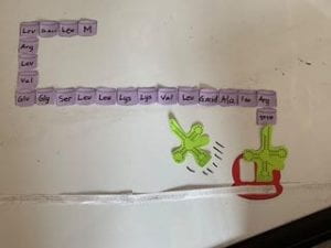
There are 2 “sites” in the ribosome: the P site, and the A site. These sites are tRNA sites and is practically a “loading zone” for tRNA molecules. The tRNA which has the amino acid Methionine, and the anticodon of UAC, will pair with the codon of AUG on the mRNA strand (see photo below). We call this the initiator tRNA, and this is always located at the P site and while that is happening, the A site is vacant and is able to be used up.

Elongation is the process of repeatedly bringing the correct amino acids with the correct anticodons to the next set of codons, in other words, this is the process of elongating the polypeptide chain. Since the A site is now vacant, the appropriate tRNA comes into the A site for its corresponding codon. For example, if the next codon on the mRNA strand is UUG, the tRNA will have an anticodon of AAC and the amino acid attached will be Leucine.

Once both sites are occupied, the tRNA in the P site will give its amino acid to the tRNA in the A site, and start a chain supported by peptide bonds between the amino acids (see photo below).

After the amino acid is passed on, the tRNA in the P site will leave, and the mRNA strand will shift along with the tRNA. That tRNA which was normally in the A site will be shifted to be in the P site now, and therefore, make it so that the A site is now vacant again. In the vacant A site, a new tRNA will be inserted with the correct anticodons and amino acid for the codon on the mRNA strand.


The same process is repeated up until a “STOP” codon is reached. This step is the termination step. Just like there is a “start” codon – Methionine – there is also a “stop” codon. There are 2 different stop codons: UAA, and UAG. However, these codons do not have a matching tRNA. So no new amino acids are able to be added to the polypeptide chain; therefore, the ribosome dissociates and the polypeptide chain is released, and the components are all split up.


The models we have been using in our class’ activity has been extremely helpful to understand how the systems work. I strongly think that visuals can be more helpful than words from text because we get to experiment with our own hands of how something works. Additionally, it was easier to understand with bigger sized components. After we figured it out, it was only a matter of shrinking everything down to actual size and still, we would know how everything would be working inside our bodies. The models were used to show the unzipping of the specific gene section of the DNA and it showed how the bonds were broken during the process by using RNA polymerase. It also showed how while the DNA strand was being unzipped, the RNA polymerase was also complimentary base pairing and creating H-bonds between the bases with the nonsense strand on the left in order. We can see that the adjacent phosphate/sugar groups are not covalently bonded yet as well and when complimentary base pairing is finished, it is apparent how the backbone is fully glued with each other and create a proper strand of mRNA. We can also identify the separation stage through the models by how the H-bonds break between the mRNA and DNA and mRNA separates itself with the nonsense strand; in the meantime, the sense strand is bonded back together with the nonsense strand with H-bonds and reverts back to its original state of DNA.
Although the models may help with understanding greatly, it does lack one of the principal factors: the demonstration of the double-helix shape. I understand that it very difficult to show with the models we have used; however, if this was a model activity for a person that was very new to the material, it would be better if the model could show all of the important features for an enhanced understanding experience. Since the model does not show the shape of a double helix, only one half of the first step of transcription is demonstrated: unwinding cannot be shown. I think another improvement can be made with the mRNA strand during translation. The improvement can be with proper distribution of the codons on the mRNA strand so that they match perfectly with the P and A sites of the ribosome. If we look at the photo below, we can see that we had to move the mRNA strand back and forth to be able to match the anticodon on the tRNA because if it was matched to one side, it would be messed up on the other.

If these features of the experiment could be improved, I think it can drastically affect the learning experience of a student because this activity would cover everything which was learnt in this course about the structure, and functions that specific parts of the cell perform. And since the models are enlarged into life-size models, it helps with understanding how the specific parts of the cells function inside our bodies and we can see their exact processes they go through.
I think models are the best method to demonstrate how things work and how they appear. Sometimes models may be overlooked due to their sheer simplicity of appearance and functionality. To non-scientists, there may be mixed opinions; either some thinking that it’s super easy to understand and finding it very beneficial, or some people that think of stereotypes of scientists that they’re always very complex with their work, and that the models do not show the professionalism of a true scientist. However, through this modelling activity, I have learnt that simplicity is often very profitable in terms of knowledge of specific functions and the processes. I found that it was very easy to understand with the models because it was a hands-on activity and I was personally moving things around and saw how specific components worked. As an individual that is still yet very fond with biological sciences, I can second that it is a very effective way to educate the public about science as well. Despite having access to notes beforehand and subsequent lectures, I think that as someone that doesn’t know any prior information could have learnt so much from this modelling activity alone because it was very clear and easy to understand. Although this may be simply a personal comment, but I think that sometimes people learn best when they are showed a concept in the simplest ways and elaborating on the concepts by showing your understanding with the use of physical models because it is possible to figure things out easier when working with your hands.
In general, I think that our modelling activity was pretty close to being immaculate. We took photos during the process that can prove our impeccable procedures, and it was exactly up to par with the given instructions and how the instructions wanted us to execute the activity. Therefore, I think I learned a lot through the use of these models and I can guarantee these models can easily help someone else as well.
References:
- A+P12 P5 Fall 2022 Notebook (Cell Function, DNA ProteinSynthesis, Protein Synthesis – transcription)
- A+P12 P5 Fall 2022 Notebook (Cell Function, DNA ProteinSynthesis, RNA Transcription Model)
- A+P12 P5 Fall 2022 Notebook (Cell Function, DNA ProteinSynthesis, Translation)
- A+P12 P5 Fall 2022 Notebook (Cell Function, DNA ProteinSynthesis, Translation steps)
- https://humanbiology.pressbooks.tru.ca/chapter/5-6-protein-synthesis/
September
9
Magnetic Poetry

September
8
