DNA (Deoxyribonucleic acid)
Structure of a DNA (Deoxyribonucleic acid)
The structure of DNA is made up of 3 main components. Sugar and phosphate groups making up the 2 “backbones” and nitrogenous bases. The sugar-phosphate on each “backbone” are antiparallel structured, meaning that the phosphates on one “backbone’ reads in one direction and are above the sugar group, while the other “backbone” reads in the opposite direction and the phosphate is below the sugar group. The nitrogenous bases are attached to the sugar group and another nitrogenous base through H-bonding. Each nitrogenous base has a complimentary base pairing. If Guanine (G) is present on one “backbone”, then Cytosine (C) must be connected on the other “backbone”. Same thing happens with Thymine (T) and Adenine (A).
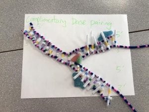
(Section of the nucleotide H-Bonded with their complimentary base)
(Pink = Phosphate, Yellow = Adenine (A), Blue = Thymine (T), Green = Cytosine (C), Purple = Guanine (G), Blue pipecleaner = “backbones”, White pipecleaner = hydrogen bonds)
How does this activity help model the structure of DNA? What changes could we make to improve the accuracy of the model?
This activity helps understand the basics of modeling a DNA molecule down to larger pieces, which means that showing all bonds within the molecule aren’t necessary. That makes the activity less tedious and time consuming. A way to improve this activity would be to expand the size of the DNA molecule in order to show how the sugar bases, nitrogenous bases and phosphate groups are bonded together. This can be shown by creating each section of a nucleotide separately, then combining them with a couple other nucleotide together to make a “zoomed in” model of a DNA molecule.
DNA replication
When does DNA replication occur?
DNA replication happens when cell occurs. It will allow all new cells to have the same DNA across all the new and old cells.
Name and describe 3 steps involved DNA replication. Why does the process occur differently on the “leading” and “lagging” strands?
- Unwinding and Unzipping: The double helix DNA molecule is unwound into a anti-parallel strands, then unzipped by the enzyme “DNA helicase” from the bottom of the molecule.
- Complementary Base Pairing: Next, the enzyme DNA polymerase attaches to a leading strand and a lagging strand, one for each. The leading strand is read from “bottom to top”, 3′ to 5′, meaning that the polymerase would pair the complementary bases to one another constantly until the entire molecule is paired. The lagging strand is read from “top to bottom”, 5′ to 3′, meaning the polymerase would pair complementary bases in sections in order to pair up all nucleotide in the molecule, since the helicase is constantly unzipping more nucleotide behind the lagging polymerase.
- Joining: The nucleotide on the new strands covalently bond to it’s new partner via DNA ligase. After all nucleotide on both the leading and lagging strands are bonded, the 2 new daughter molecules are successfully duplicated and are wound back up into the double helix shape.
Since each “backbone” can only be read in one direction, the DNA polymerase can only read that strand in the direction it’s given. One DNA polymerase would read towards the helicase where the DNA is being unzipped, while the other DNA polymerase would read away from it, meaning after a certain point, it would have to travel backwards to facilitate the complementary base pairings to the newly unzipped nucleotide.
What did you do to model the complimentary base pairing and joining of adjacent nucleotide steps of DNA replication. In what ways was this activity well suited to showing this process? In what ways was it inaccurate?
We had the blue bigfoot candies represent a DNA polymerase, red bigfoot represent DNA ligase, and a watermelon slice represent the helicase. The blue bigfoot sat on top of the DNA molecule to show that it undergoes pairing. While the red bigfoot sat on top of the DNA molecule as well showing the joining of the complementary base pairs.
(Original DNA with replicated DNA)
This model helps accurately show the basics of what happens to DNA as is undergoes replication. The candies help show an introduction to where the specific enzymes sit as they do their jobs.
The model doesn’t show how the enzymes do their jobs when DNA replication occurs.


