1- how is mRNA different from DNA ?
– DNA is made up of deoxyribose sugar while mRNA is made up of ribose sugar.DNA has thymine as its pyrimindine base while mRNA has uracil as its pyrimidine base. DNA is present in the nucleus while mRNA diffuses into the cytoplasm after synthesis. DNA has 2 strands that are anti-parallel that intertwine to form a double helix while mRNA is short and single stranded. mRNA is short lived , while DNA has a long lifespan.
2- describe the process of transcription
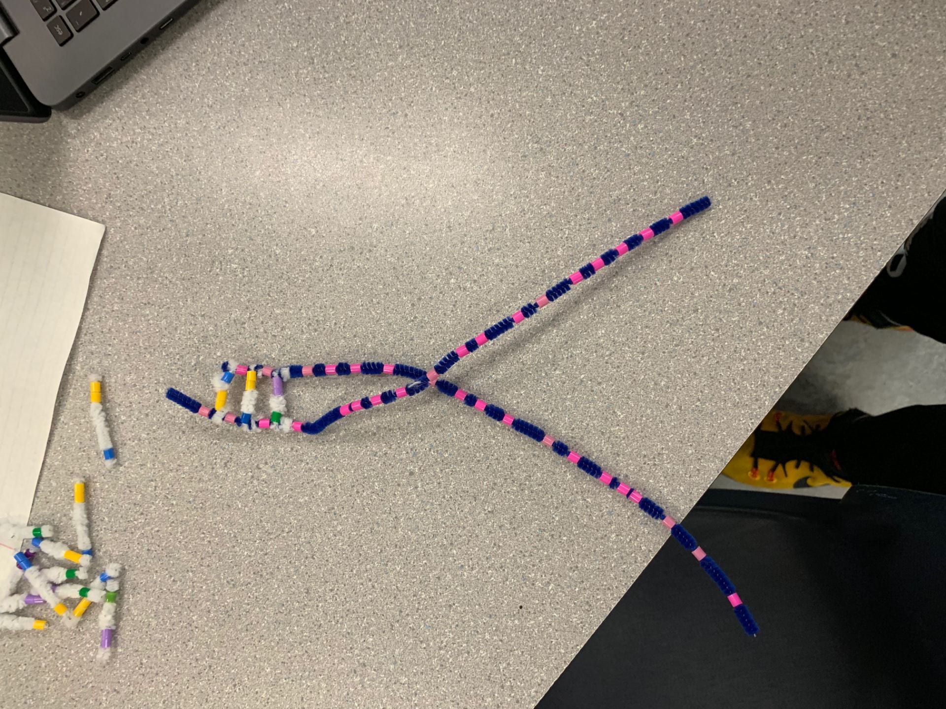
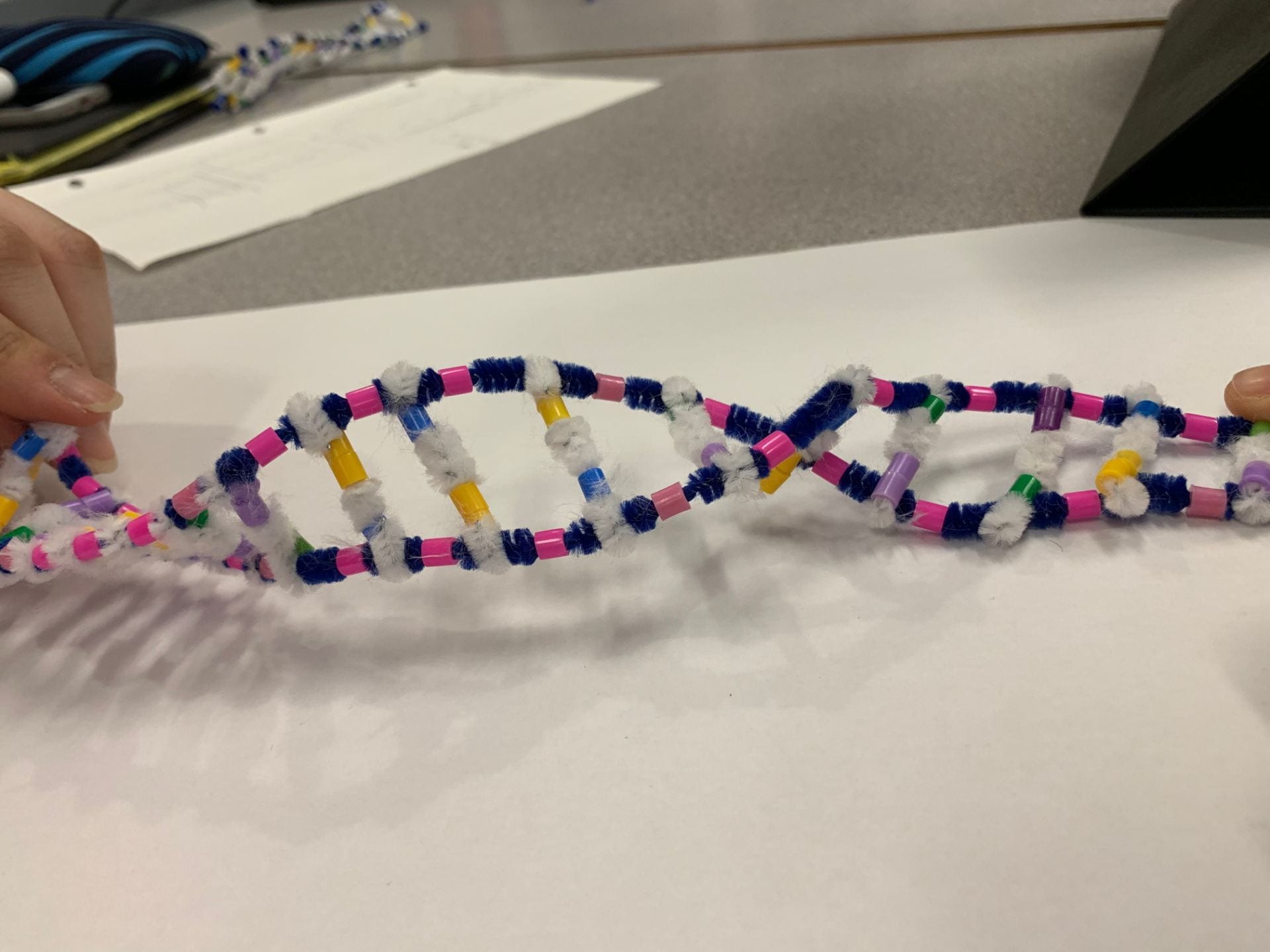
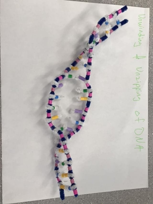
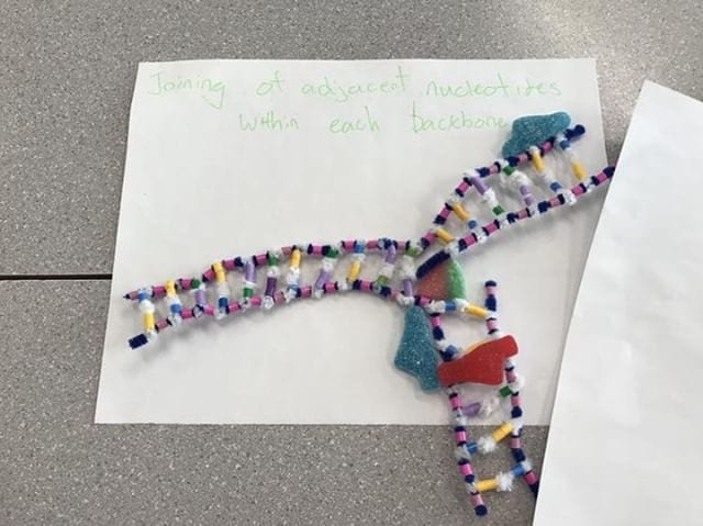
the process of transcription begins with a specific section of the DNA unwinding which exposes a set of bases. Along the sense strand of DNA complementary RNA bases are brought in. In RNA , uracil binds to adenine . While in DNA cytosine binds to guanine . In the other strand of DNA ( missense strand) is not read read in eukaryotic cells.Adjacent RNA nucleotides form sugar – phosphate bonds and then the RNA strand is released from DNA . After that the DNA molecule rewinds and returns to its double helix form. This process occurs in the nucleus and is facilitated by RNA polymerase which is depicted in the form of a fuzzy peach in the photo below . The photo shows the RNA polymerase copying a particular segment of DNA into RNA .after this is all done the red RNA strand separates from the DNA strands .
 RNA is shown as the red strand
fuzzy peach – RNA Polymerase
3- how did today’s activity do a good job of modelling the process of RNA transcription? In what ways was our model inaccurate .
– The Pipe cleaner model is an excellent way to learn the steps of transcription because it is hands on and helps us better understand the process since we are building it ourselves . In addition to being hands on , the activity allows us to break down transcription into several steps and observe the process bit by bit and take our time to understand each step clearly. The different strand colours made it easier to distinguish between DNA and RNA and gave me a better understanding of the entire process rather than just reading the lesson of a board. The beads make the bases visually appealing and easier to distinguish , especially between single or double ringed bases.On the other hand , some discrepancies lie within this lab. For starters , the process of transcription is continuous meaning it happens without any stops or breaks. Another innaccuracy is with the size of the strands . Normally the DNA strands are much longer than the RNA but as you can see of our picture the red strand is almost as long as the DNA strand which in reality is false since RNA is much shorter.
Translation Activity
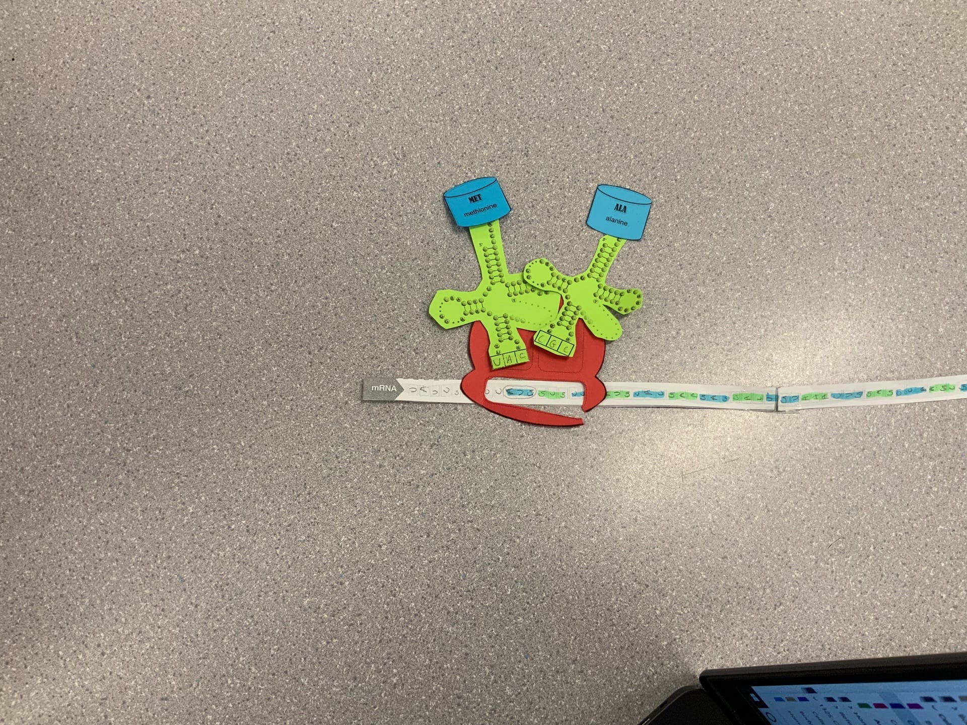
1- describe the process of translation- initiation , elongation , and termination. ( photos illustrate the process in order)
– initiation: The process starts with the mRNA reading the start codon (AUG) and attaches to the R site of the ribosome . The AUG codon always initiates translation and codes for the amino acid : methionine . tRNA binds to the start codon of mRNA . The tRNA has a binding site of 3 bases called anticodons that are complementary to the mRNA codon. The methionly-tRNA is in the P site of the ribosome . The A site next to it is available to the tRNA carrying the next Amino Acid.
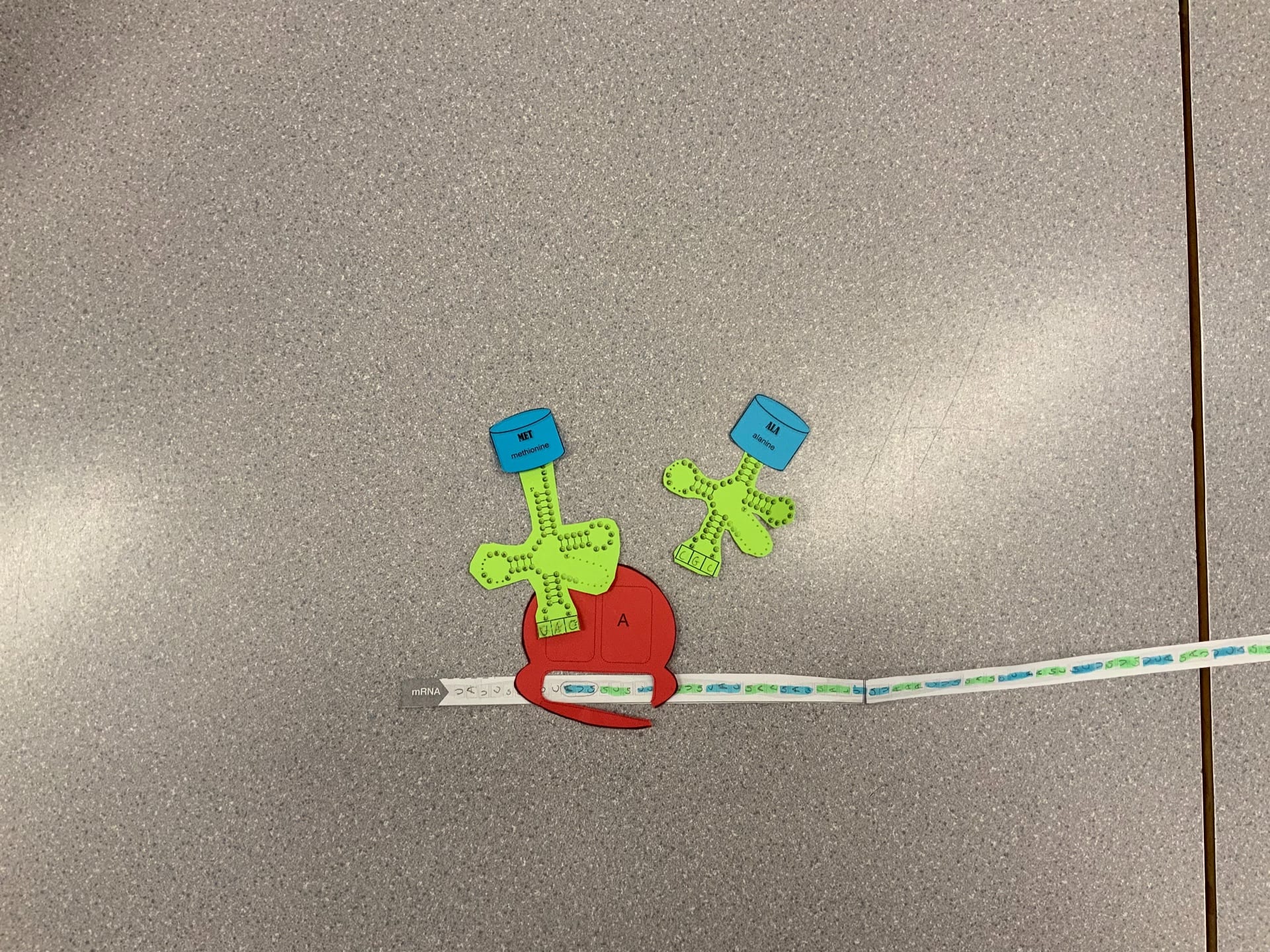
– elongation : more amino acids are added and connected together to form a polypeptide. A Peptide bond is formed between the new amino acid and the growing polypeptide chain. Then , the amino acid is removed from the first tRNA ( bonds break).
The first tRNA that was in the P site is released , and the tRNA in the A site is moved over to the P site.
The ribosome moves over one codon along the mRNA . This movement shifts the second tRNA to the P site . The third tRNA with the third amino acid can move into the A site and bind with the next codon on mRNA.
the process repeats and the chain elongates .
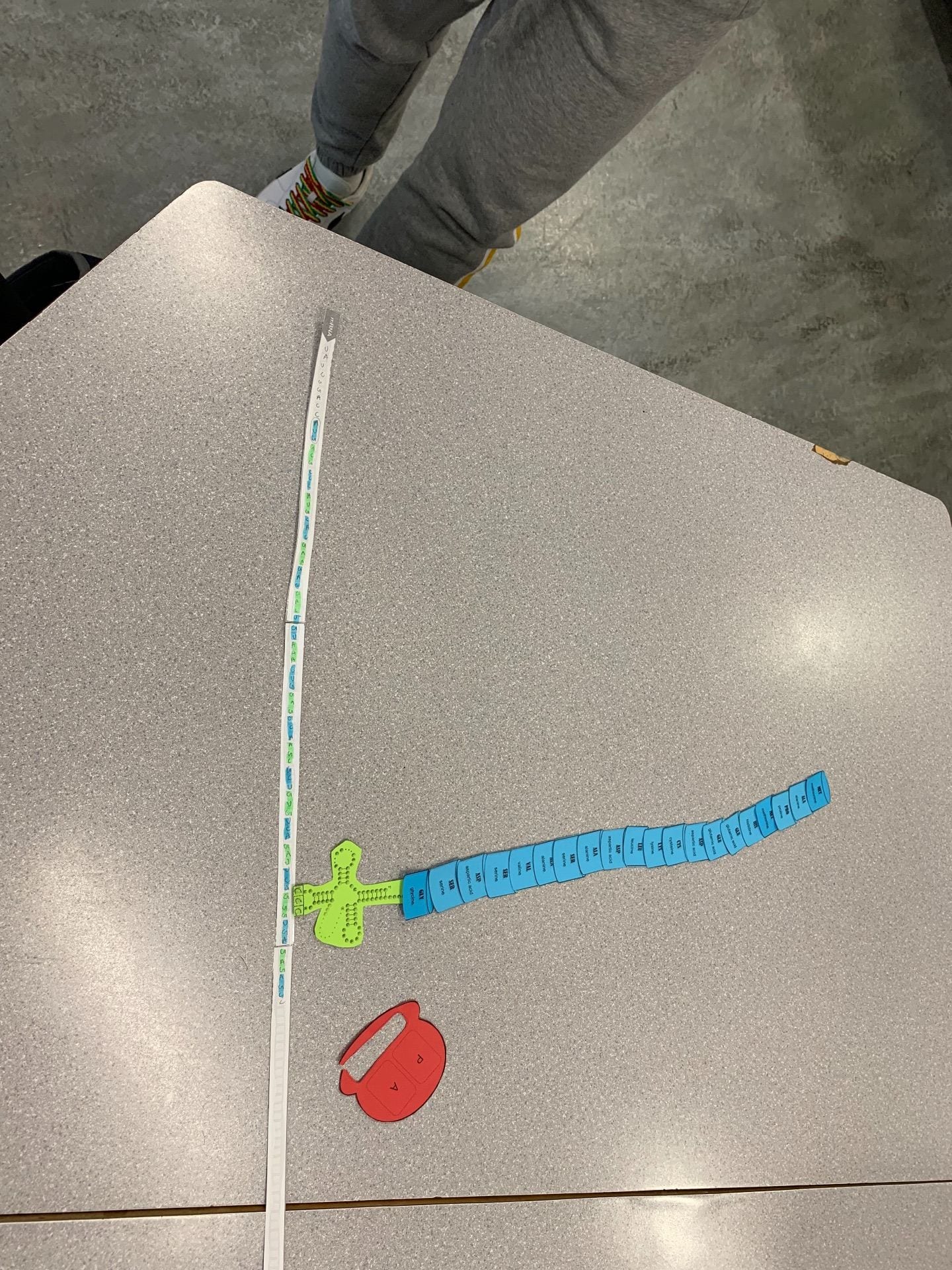
termination :the elongation process repeats until the stop codon is reached. ( UAA , UAG , UGA)
the stop codons do not code for amino acids but act as signals to stop translation
A protein called release factor binds directly to the stop codon in the A site. This causes a water molecule to be added to the end of the polypeptide chain , and the chain then separates from the last tRNA.
The protein is complete now. The mRNA breaks down and the ribosome splits into large and small subunits .
The pictures below illustrate the three steps of translation : initiation, elongation , and termination .
2- how did today’s activity do a good job of modelling the process of translation? In what ways was it inaccurate?
The activity did a good job of modelling translation by breaking it down into several steps and showing each small step for us to fully grasp the concept of the A and P sites , tRNA anticodons , and how translation begins. the different paper figures and colours made the process visually easier to grasp and understand. The fact the activity was hands on made it more meaningful and easier to understand. On the other hand , it was not possible to show the 3 D components of the process as this process is usually happening continuously and at a steady pace. In the end , I believe this activity gave me a better understanding of translation and definitely helped when I went back and read the steps of translation. Since we worked hands on , I better understood the process and re reading the lesson helped reinforce my understanding p.


–

 RNA is shown as the red strand