Powerpoint 1 (General): March 1 Kingdom Protista
Powerpoint 2 (Ciliophora and Zoomastigina): March 7 Kingdom Protista
Powerpoint 3 (All phyla): March 3 Animal-like Protists
- Explain how structure, function, environment and cost-benefit are related (Game)
- Explain the current theory on how Protists evolved from Monerans
- Identify and Label the generic Protist structure
- Identify the structure and functions of Paramecium parts
- Describe reproduction of Paramecium
- Compare and contrast the Protist Phyla
- Provide examples of Protist Phyla
-


Kingdom Protista is a group created by exclusion. Historically, taxonomists and biologists alike had much trouble classifying these organisms. The reason for this difficulty is that members of Kingdom Protista have characteristics not unlike Animalia, Plantae or Fungi. Unlike the other four kingdoms (Animalia, Plantae, Fungi and Monera), which are relatively clear cut, members in Kingdom Protista are very diverse and share little in common. Lynn Mangulis, the scientist who came up with the endosymbiont hypothesis, wrote that the Kingdom Protista
“is defined by exclusion: its members are neither animals…, plants…, fungi …, nor prokaryotes.”
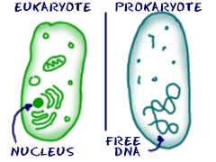
Perhaps a more scientifically sound definition: “unicellular (single-celled) organisms that are eukaryotic”. As you will recall from our taxonomy unit, Prokaryotes are organisms without a nucleus or membrane-bound organelles, and Eukaryotes are organisms with a nucleus and membrane bound organelles. (Ribosomes are organelles, but are not membrane-bound)

http://study.com/academy/lesson/eukaryotic-and-prokaryotic-cells-similarities-and-differences.html

Therefore, a protist is simply a unicellular organism with:
- Nucleus containing DNA
- Membrane bound organelles
Which about sums up all that these organisms have in common.
But where did these organisms come from? Where did they evolve from?
Several observations seemed to provide some clues as to how it happened.
- Mitochondria and Chloroplasts, organelles that exist in eukaryotic cells, have their own DNA. The DNA is completely separate from the DNA of the eukaryotic cell itself.
- Some Protists have organelles that can be removed, without ill effect on the Protist. Some of these organelles can even grow on their own!
- Some of the Eukaryotic cell’s organelles are structurally, very similar to Prokaryotes
Noticing these characteristics, Lynn Margulis proposed the Endosymbiont Hypothesis. The hypothesis states that organelles are actually descended from prokaryotes that lived inside another prokaryote in a symbiotic relationship. Each benefited the other. For example, a blue-green bacteria that lived in a bigger moneran had shelter, while the bacteria provided the host cell with sugars and nutrients. At some point, the blue-green bacteria may have lost their independence, and became the precursors to the organelles we see today.
A creative way of explaining Endosymbiosis.
Paramecium sp. (Phyla: Ciliophora)
Paramecium is a model organism for protists. Like most model organisms, it has been studied extensively and is easily cultivated (grown). However, a model organism is not necessarily representative of the group it comes from. Just as a lab rat is not necessarily representative of all mammals, or even all rodents, so we should not assume that Paramecium is a perfect representation of Protists.
Movement | Paramecium sp. is classed in Phylum Ciliophora, a protist phylum characterized by cilia.
Feeding | Paramecium sp. feeds on small organisms, such as bacteria. The cilia first sweep the food toward the oral groove, a small opening where food is trapped. The oral groove leads to the gullet, which produces food vacuoles, a small sac where food is stored. the food vacuole then contacts lysosomes, organelles containing digestive enzymes. These digestive enzymes then break down the food, and the wastes are expelled from the anal pore.
Water Balance | Paramecium sp. live in freshwater environments. Because the Paramecium sp. has a higher solute concentration than the freshwater around it, water tends to move inside the cell. Therefore, excess water must be expelled from the Paramecium sp. from the cytoplasm to the contractile vacuole.
Reproduction | Paramecium sp. is able to reproduce via binary fission (asexual reproduction). The micronuclei are duplicated, and the macronucleus is split apart. The split occurs lengthwise, like pulling silly putty apart. During conjugation, however, two paramecium arrange side by side and exchange genetic information.
No new daughter cells result from conjugation, therefore it is not a form of reproduction. However, the exchange in genetic information does lead to an increase in genetic diversity amongst the population. An increase in genetic diversity leads to more diverse traits, and having more traits means there’s a greater chance that at least a few of the cells may have the correct traits to survive, even thrive, in the environment.
Therefore, asexual reproduction occurs more often in stable, unchanging environments. Under these conditions, because Paramecium sp. are likely to survive and thrive, energy is better invested in increasing population numbers. However, all offspring will be genetically identical.
Conjugation occurs more often in unstable, changing environments that are stressful. Even if Paramecium sp. numbers increase, because the genetic diversity is low, there is less likely to be individuals that have the gene combination to help them survive in the environment. therefore, it is much more beneficial for Paramecium sp. to invest their energy in conjugation, where gene combinations are reshuffled.

(Binary Fission)
(Conjugation)
Kingdom Protista Phyla
We cover nine Protist phyla in this course: 4 animal like, 3 plant like and 2 fungi like.
|
Animal-like Protists (Heterotrophic) |
Plant-like Protists (Autotrophic) |
Fungi-like Protists (Heterotrophic) |
| Ciliophora
Zoomastigina Sporozoa Sarcodina |
Euglenophyta
Pyrrophyta Chrysophyta |
Acrasiomycota
Myxomycota |
| Phyla | Picture | Distinguishing Characteristics | Examples |
| Ciliophora
“Ciliates” (“Cilia-bearer”) |
 |
Animal like protist with cilia | Paramecium sp.
Stentor sp. Vorticella sp. |
| Zoomastigina
“Zooflagellates” |
 |
Animal like protist with flagella | Trypanosoma
Trichonympha |
| Sporozoa |  |
Non-motile, spore producing animal like protist | Plasmodium |
| Sarcodina |  |
Pseudopods
Some produce silicon shells (SiO2) |
Ameba Heliozoans Foraminifers Radiolarians |
| Euglenophyta |  |
Flagellum
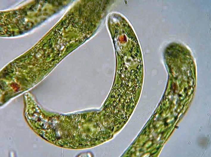
Chloroplasts and red eyespot Structurally similar to zoomastiginan Phototrophic Heterotrophs |
Euglena sp. |
| Pyrrophyta
Dinoflagellates (“fire-plant”)
|
 |
2 flagellum, one wrapped around like a belt.
Thick plates, armored appearance Named for bioluminescence in some members of this phyla. |
Gonyaulax polyhedron (Red tide)
|
| Chrysophyta
(“golden plant) |
 |
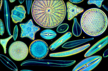
Beautiful silicon shells
Cell walls made out of pectin Stores food as oil rather than starch to stay afloat. |
Diatoms |
| Acrasiomycota |  |
Cellular Slime Molds
Lives part of life as amoeba Individuals aggregate (group together) into one mass and migrate when food runs low. Produces fruiting bodies that spread spores. |
|
| Myxomycota |  |
Acellular slime molds
Life part of life as amoeba. Produces plasmodiums, a large single celled, multinucleate mass that may stretch a few centimeters. Produces fruiting bodies that spread spores |
Dog vomit |

