Microscopes

Compound microscopes are a basic tool of biology. They are standard for a number of fields, including health care, research, toxicology, and many many more.

They are also just really cool in general.

compound-microscope-parts-diagram

Parts of a Microscope 

  1. Ocular Lens – has a power of 10x
  2. Objective Lens(es) – a set of lenses with three or four different powers
    1. Low power (4 x)
    2. Med power (10 x)
    3. High power (40 x)
  3. Stage – where the slides are placed
  4. Diaphragm – a dial that can be turned to control the amount of light coming through
  5. Coarse focus knob – shifts the height of the stage to help focus. As the name suggests, the coarse adjustment knob shifts the stage faster and should be handled with caution
  6. Fine focus knob – shifts the height of the stage to help focus, but very very slightly.
  7. Light 

Total Power 

The total magnifying power of a microscope is calculated like this:

Ocular Lens Power  x   Objective Lens Power  =   Total Lens Power

In the case of our microscopes, the magnifying power of the ocular lens is 10x. The magnifying power of the objective lens vary (4 x, 10 x, 40 x).

Therefore, the total magnifying power at all levels is:

Low     10  x   4   =   40 x
Med    10   x   10 =   100 x
High   10   x   40 =  400 x

Creating a Wet Mount 

  1. Obtain a clean slide and coverslip (be very careful when handling these as both are glass)
  2. Put your specimen flat onto the slide.
  3. Put a drop of water onto your specimen
  4. Put the coverslip on. Make sure to put it down at a 45 degree angle. This pushes out the air to reduce air bubble formation
  5. If there is too much water on your slide, gently dab away with a paper towel.

Calculating size of specimen based on the field of view  

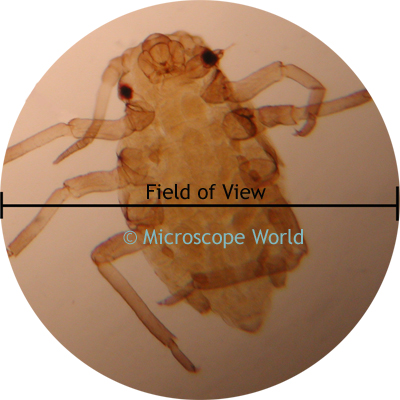
Suppose you saw this flea under your microscope. How could you figure out how large it really is? One critical piece of information is just how wide is the field of view? 

When we are looking at low power, the field of view will be wider than if we are looking at high power, since we are now focused on a smaller area.


At Low Power, the field of view is about 4.2 mm, or 4200 um (1 mm = 1000 um)
At Med Power, the field of view is about 4.2 mm, or 1680 um (1 mm = 1000 um)
At High Power, the field of view is about 4.2 mm, or 420 um (1 mm = 1000 um) 

So how do we calculate the field of view? Suppose we were looking at the cells above at medium power and I was trying to figure out how big the cell is.

I know that the diameter of the field of view is 1680 um. And I estimate that about 6 cells fit across the diameter.

Therefore

1680 um / 6 = 280 um

This means that the cells are about 280 um across.

Biological Drawings Rubric

CCI03032016 

Kingdom Monera

February 25 Powerpoint: February 25 Kingdom Monera
February 29 Powerpoint: February 29 Kingdom Monera (cont’d)

Learning Objectives 

  • Identify and label structures of a generic Moneran
  • Identify and Describe the four criteria through which Moneran are classified
  • Describe the ways in which Moneran obtain/metabolize energy
  • Describe the three ways Moneran reproduce

Kingdom Monera is a large, diverse and wholly under-appreciated group of organisms. They perform many critical functions in the ecosystem, such as detritivores. They are used in the food industry for production of foods, such as milk or cheese. They are also responsible for many human diseases, such as the black plague that destroyed a third of the European population in the 14th century.

Members of this kingdom are also called bacteria.

Generally, bacteria all share some common features.

Structure of generic Monera 

Picture3


  • All Monera are unicellular and prokaryotic
  • No nucleus. DNA or RNA and ribosomes floating in cytoplasm.
  • Cell membrane made of lipids
  • Cell wall made of peptidoglycan to protect cell
  • Flagellum (plural: flagellato move


Ways to classify a Moneran

Because of their small size, bacteria are very hard to observe under the microscope. Even when their individual shape or form can be seen, many look far too alike to definitively classify. Thankfully, we do have ways to classify Monera.

  1. Shape – Almost all Monera fit into one of three shapes: round and spherical (cocci), long and rod shaped (bacilli) and spiral shaped (spirilla). Cells can also be classified based on their clustering behavior. Some cluster into colonies and strings, while others are primarily solitary.
  2. Gram stains – because bacteria are for the most part, transparent to the naked eye, they must be stained to be properly observed under the microscope. When Hans Christian Gram, a Danish bacteriologist was staining bacteria with crystal violet, he realized that some bacteria retained the stains and became purple, while some did not and became pink. As it turns out, this has to do with the thickness and structure of the peptidoglycan cell wall. Generally, a thicker cell wall will retain more of the crystal violet, while thin ones will not.
  3. Bacteria movement – some bacteria have flagella, whip like projections, that allow them to move. Some bacteria secrete slime to move along like a slug. Still others don’t move at all.
  4. DNA/RNA Sequencing – the most specific way to identify a bacteria is probably through DNA/RNA sequencing. We do so by identifying the genes of the bacteria, which is probably fairly specific to each species. This method has become more common in recent years as the cost of genome sequencing decreases.

Ways Moneran get their energy

It should be noted that the following terms describe where organisms (any organism, not just bacteria) get their energy from.

  1. Phototrophic Autotroph: organisms that derive their energy from the sun
  2. Chemotrophic Autotroph: organisms that derive their energy from inorganic chemicals (chemicals that are not derived from living things)
  3. Chemotrophic Heterotroph: (aka heterotrophs) are organisms that derive their energy from organic chemicals (chemicals that are derived from living things)
  4. Phototrophic Heterotroph: are organisms that derive their energy from organic chemicals or the sun, depending on which source is available

————- (End of Thursday, February 25) ——————-

Ways Monerans Metabolize their energy 

Depending on whether the Moneran uses one or both of the following

1. Cellular Respiration – a series of chemical reactions that uses sugars and oxygen to produce energy.
2. Fermentation – a series of chemical reactions that converts sugars to acids, gases or alcohol, and produces energy. It occurs without oxygen.

we classify monerans as follows.

  1. Obligate Aerobes: Monerans that need oxygen in order to produce energy (aerobic respiration). These Monerans mainly use cellular respiration.
  2. Obligate Anaerobes: Monerans that will die in the presence of oxygen, as oxygen is toxic to these monerans. These Monerans mainly use fermentation.
  3. Facultative Anaerobe: Monerans that can produce energy in oxygen (cellular respiration), but switch to fermentation if there is no oxygen.

Ways Monerans Reproduce 

  1. Binary fission – where the cell replicates its DNA, and splits into two daughter cells, where each daughter cell gets one set of DNA
  2. Conjugation – the bacterial form of sexual reproduction. Involves the exchange of genetic information.
  3. Spore Formation – strictly speaking, not a form of reproduction. Occurs when a bacterium produces a hard covering (spore) and becomes metabolically inactive to preserve itself in adverse environments.

https://www.youtube.com/watch?v=EtxkcSGU698

Bacteria Research Mini-Project – One page fact sheet on an assigned bacterium – DUE TUESDAY FEBRUARY 1st 

  1. Block A: https://docs.google.com/document/d/1DxJICGG2N96HHj9MwghvtcQ0w1mLOn0UfuBRhlUVdUA/edit?usp=sharing
  2. Block C: https://docs.google.com/document/d/1WN7R50RWj-oKsa0CoJetRjI7q-kFhzu0ZAYVru5FZVQ/edit?usp=sharing

Viruses

Powerpoint: February 22 Viruses

Keywords: Virus, head, tail, DNA, RNA, protein capsid/coat, lipid membrane (or membrane envelope), antigens, lytic cycle, lysogenic cycle,  

Textbook Pages: 17-1

Learning Objectives 

  • Identify the structures of bacteriophages and retroviruses
  • Describe the life cycle of (1) Lytic and (2) Lysogenic viruses
  • Describe the body’s defence mechanisms against a viral/bacterial infection

What are Viruses? 

Viruses are defined as “noncellular particles made up of genetic material and protein that can invade living cells”. They do not belong to any of the five kingdoms of organisms. It isn’t even clear whether viruses are living or non-living. While they do evolve in so much as only successful viruses can propagate their genetic information, they do not have cells, do not grow or reproduce, and do not use energy the way living things do. They also cannot live independently of cells, as they depend on cells to replicate.

Nevertheless, viruses are fascinating tiny machines of nature that have been used and studied for a variety of purposes, such as in genetic engineering, combatting cancer, and to destroy harmful bacteria in foods. Some viruses are also responsible for some of the most debilitating diseases known to man, including HIV (Human immunodeficiency virus) leading to AIDS, mono, shingles, herpes and chickenpox.

Identifying Viruses 

Viruses come in many shapes, sizes and life cycles. There are two specific groups of viruses we will focus on here.

Bacteriophages

Retroviruses

phage-21

Bacteriophages have a distinct shape and structure.

There are two distinct parts: a head and a tail. The head is composed of a protein capsid or coat. It contains the DNA information. The tail and fibers at the base are used to attach the virus to bacteria, where it can then inject its genetic information into the cell.

retrovirus 2

Retroviruses are named for their ability to insert their genetic material into the existing genetic information of the host cell.

Normally DNA is “transcripted” to RNA (nucleic acid made of a single chain of nucleotides, in contrast to two strands in DNA). However, the genetic information in retroviruses are made up of RNA.

When the RNA enters the host cell, enzymes attach the piece of RNA to the DNA of the cell. Complementary nucleotides then attach to the RNA, making it double stranded and indistinguishable from host cell genome.


Just like the head of bacteriophages, made of a protein capsid coat with genetic information inside, retroviruses a protein capsid with RNA. However, they are also surrounded by a lipid membrane (membrane envelope) and often antigens.

Antigens are structures on the outside of the virus, toxins or proteins. The antigens act like the face of a virus, which can be recognized by the body and targeted by the body.


technology

DNA Replication

DNA do not techninically reproduce the way living organisms do. However, they do invade host cells, causing them to create more copies of viruses. There are two ways that this happens.

Lytic Cycle

Lysogenic Cycle

Picture1

  1. Virus attaches to host cell
  2. DNA information is injected into the cell
  3. Cell reads the virus’s genetic information (it cannot tell the difference between its own DNA and viral DNA)
  4. Cell replicates viruses inside itself
  5. Viruses become very numerous until the cell eventually bursts releases the viruses
NOTE: Bacteriophages can also replicate through the lysogenic cycle. Retroviruses almost always replicate through the lysozenic cyccle.

Picture2

1, Virus attaches itself to host cell
2. DNA/RNA information is injected into the cell |
3. Virus Genetic information is incorporated into the host cell’s genetic information, where it remain dormant for many years.
4. As the cell goes through cellular respiration, each daughter cell will also contain virus genetic information
5. At some point, the viral genes are activated, and replication begins.
6. Again, the viruses become very numerous until the cell bursts.


Thankfully, the human body also has some ways of protecting ourselves against viruses, at different levels of specificity.

 Primary Line of Defense: 

These forms of defense are non-specific, as in, it is to protect against any form of pathogen, virus, bacterial, protist, etc.

– Skin

– Oil and Sweat

– Hairs and cilia in mouth and nose

– Stomach (acids)

– Saliva, sweat and tears (lysozyme)

 Secondary Line of Defense: 

These forms of defense are also non-specific, but are generally activated only when pathogens have invaded.

– Inflammatory response: white blood cells

– Fever

 Tertiary Line of Defense 

The most specific form of defense. These forms of defense are activated specifically to target the pathogen.

This is usually done through antibodies that are produced based on the antigens on the surface of the pathogen. The antibodies can then target the pathogens.

Interferons : produced by cells to interfere with virus replication

–  Antibody production: white blood cells produce antibodies