Self Assessment on Oil Change Lab
Author Archives: Kiera
Chemistry 12 – Self Assessment
Chemistry 12 – Lab on Acids and Bases
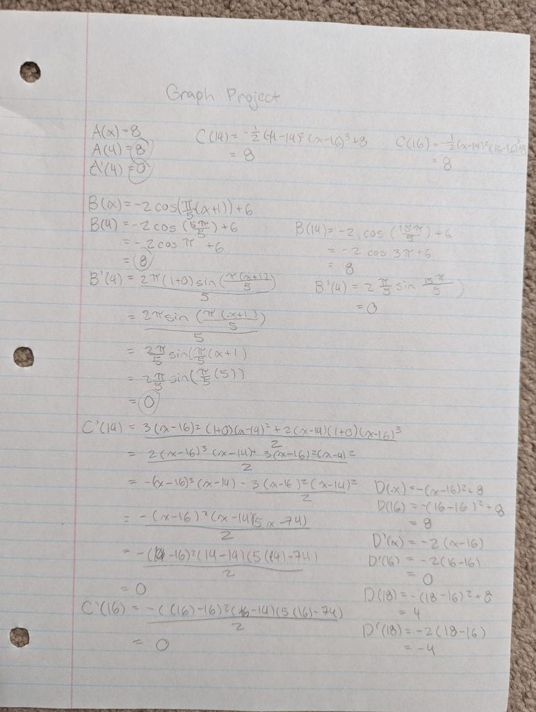
Self Reflection – Desmos Project Calculus 12


Anatomy and Physiology 12 – DNA Replication and Protein Synthesis
DNA Replication and Protein Synthesis Lab
DNA Replication:
Figure 1: Strand of DNA

Figure 2: Model of DNA Replication

- Explain the structure of DNA – use the terms nucleotides, antiparallel strands, and complementary base pairing.
DNA is made up of two strands of nucleotides which are held together by hydrogen bonding. In between the strands are complementary base pairing, and DNA has antiparallel strands, meaning the strands each run from 5′ to 3′ and run in opposite directions from one another.
- When does DNA replication occur?
DNA replication occurs before cell division because it is required for the cell to properly divide.
- Name and describe the 3 steps involved in DNA replication. Why does the process occur differently on the “leading” and “lagging” strands?
The first step, unzipping, is when the DNA unwinds and the two strands of DNA separate.Then the next step, complementary base pairing, is when each strand of DNA is paired up with new nucleotides. The final step is adjacent nucleotide bonding, which is where the nucleotides on new strands form covalent bonds, binding together side by side with the sugar-phosphate backbone. The process occurs differently between the “leading” and “lagging” strands because they are anti-parallel to each other, meaning that one strand is the leading strand while the other is the lagging strand. New DNA can only be synthesized in a 5’3’ direction, and the leading strand has a 5’3’ direction which makes the replication of DNA molecules from the leading strand faster and continuous. Because the lagging strand goes in the 3”5” direction, DNA on the lagging strand is synthesized in short, separated segments.
- Today’s modelling activity was intended to show the steps involved in DNA replication. What did you do to model the complementary base pairing and joining of adjacent nucleotides steps? In what ways was this activity well suited to showing this process. In what ways was it inaccurate.
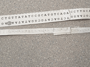
We made sure to check that the bases were correctly paired and also that the sugar-phosphate bonds were corrected drawn on the whiteboard. We also drew dashes between the base pairs that represented the hydrogen bonds that were formed. This activity was well suited to showing DNA replication because it displayed the complementary base pairing that occurs as well as the unzipping during replication, giving us a broad understanding of the process of replication. Some ways the model was inaccurate was that the enzymes such as DNA ligase were not shown in their proper shape and were instead the shapes of basic shapes instead. Also, we were not able to demonstrate the double-helix shape that DNA took.
RNA Transcription:
Figure 3: RNA transcription

Figure 4: mRNA strand

- How is mRNA different than DNA?
DNA contains the sugar deoxyribose, while mRNA contains the sugar ribose. DNA is a double-stranded molecule, while RNA is a single-stranded molecule. DNA and mRNA base pairing are slightly different since DNA uses the bases adenine, thymine, cytosine, and guanine while mRNA uses adenine, uracil, cytosine, and guanine. While DNA is responsible for storing genetic information, mRNA directly codes for amino acids and acts as a messenger between DNA and ribosomes to make proteins. While DNA completes all of its functions staying in the nucleus, mRNA’s smaller size allows it to travel from the nucleolus to the cytoplasm. Also, DNA replicates through DNA polymerase while mRNA is made through transcription using RNA polymerase.
- Describe the process of transcription.
A section of DNA unzip, exposing the bases and complementary RNA nucleotides bond with bases on the exposed strand of DNA. For RNA, uracil is the complementary base that bonds with adenine instead of thymine. The adjacent sugars and phosphates form bonds to create a strand of mRNA. The DNA then rewinds, and the mRNA is now ready to leave the nucleus for translation.
- How did today’s activity do a good job of modelling the process of RNA transcription? In what ways was out model accurate?
I found that this activity was effective in demonstrating the process of RNA transcription by showing how mRNA is created from the template of one of the DNA strands and showing the single backbone of RNA. It also showed the process of the RNA polymerase. However, the model did not show the DNA unzipping before the mRNA strand was created nor when the DNA reattaches itself after the mRNA is formed.
RNA Translation
Figure 5: Process of translation – Initiation

Figure 6: Process of translation – Termination

- Describe the process of translation: initiation, elongation, and termination.
Initiation is when the mRNA is connected by a ribosome that has an a-site and a p-site and when the p-site reads the START codon (AUG). the tRNA brings the corresponding amino acid to the start codon which starts the translation process. Then, in elongation, that process continues, forming a polypeptide chain that continues to grow as the a-site reads the next mRNA codons to bring the corresponding tRNA as the initial tRNA moves to the p-site. Termination is when there is a STOP codon that is read on the mRNA. The STOP codon does not code for any amino acids, causing the ribosome to let go of the mRNA and the tRNA to let go of the polypeptide chain.
- How did today’s activity do a good job of modelling the process of translation? In what ways was our model in inaccurate?
Today’s activity did a good job of modelling the process of translation by showing how the translation process depends on the codes of mRNA, including the START codon and the STOP codon. This activity provided a visual display of how the polypeptide chain forms and how the ribosome can hold two tRNA’s. However, this activity demonstrated translation happening in only one location while in reality, translation happens at multiple locations at a time along the mRNA strand.
English 12 – Visual Poem Analysis Assignment
A Poetry Analysis on “One fish, Two fish, Plastics, Dead fish”
English 12 – Community Connections Novel Project (Conflict)
What does happiness mean to you?
Course Self Assessment – Pre-Calculus 12
Psychological Lens Character Analysis Presentation
Character Analysis on Popular Mechanics by Raymond Carter
Identity Background Assignment
Backround Analysis on Anxious People by Fredrik Backman
 Loading...
English 12 – Analysis on Joe the Painter and the Deer Island Massacre by Thomas King
A Study on Prose Terms on Joe the Painter and the Deer Island Massacre by Thomas King