In class we modeled mitosis using pipe cleaners and string. Here are my photos to walk you through each stage of the process.
Materials:
white string – nucleus and cell membrane
pink string – spindle fibers
pipe cleaners – chromatin
beads – genes
Interphase:

What the cell looks like most of it’s life, before mitosis begins

DNA inside the nucleus is copied and duplicated
Prophase:

The replicated DNA molecules join together to form sister chromatids

Spindle fibers appear in the cell

The spindle fibers each attach to one of the sister chromosomes

The nucleus disappears
Metaphase:

Chromosomes line up in the middle of the cell
Anaphase:

The sister chromatids are separated and the halves move to the opposite sides of the cell, pulled by the spindle fibers
Telophase:

Spindle fibers disappear

Two nuclear membranes form around the chromatin on either side of the cell
Cytokinesis:
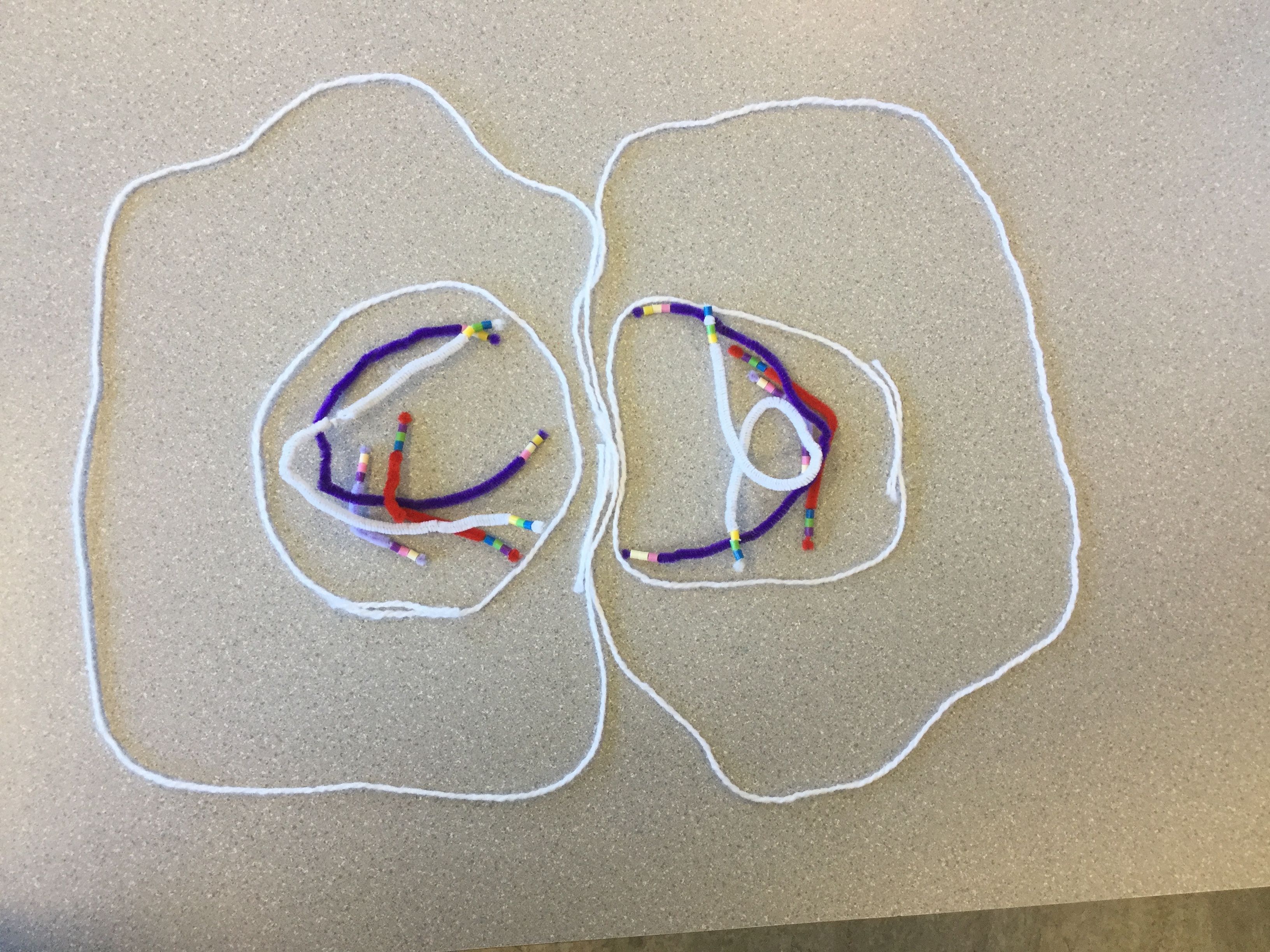

After mitosis occurs the cell divides into two identical cells
Leave a Reply